
Ovarian Cyst Management: 7 Proven O-RADS Decisions
Clinical Practice Update — Ultrasound Risk Stratification, Premenopausal and Postmenopausal Pathways, Surveillance, and Oncology Referral
This is an original clinical education article informed by current guidelines and evidence. See References below for source documents.
- Clinical Focus
- Evidence-based ovarian cyst management in premenopausal and postmenopausal women from imaging through referral
- Target Audience
- Obstetricians, gynecologists, family physicians, emergency physicians, radiologists, nurse practitioners
- Setting
- Primary care, gynecology clinics, radiology, emergency departments, gynecologic oncology centres
- Source Evidence
- •ACR O-RADS US — Ovarian-Adnexal Reporting and Data System (2020, updates 2022)
- •ACOG Practice Bulletin 174 — Evaluation and Management of Adnexal Masses (2016)
- •RCOG Green-top Guideline 62 — Management of Suspected Ovarian Masses in Premenopausal Women
- •SGO/ACOG Joint Committee Opinion — Referral to Gynecologic Oncology (reaffirmed 2020)
- •IOTA Simple Rules and ADNEX Model — External Validation Studies
Key Clinical Takeaways
Effective ovarian cyst management depends on three things: a good-quality transvaginal ultrasound, a structured risk score (ideally O-RADS), and the discipline not to operate on cysts that will resolve on their own. The rules below turn that principle into an actionable workflow.
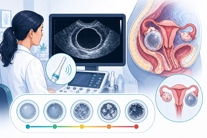
- 1Use transvaginal ultrasound as the first-line imaging modality for any suspected ovarian cyst or adnexal mass → Evaluation
- 2Assign an O-RADS score (1–5) to every adnexal lesion to make risk explicit and management reproducible → Risk Stratification
- 3Most simple cysts under 10 cm in premenopausal women resolve spontaneously — observe rather than operate → Premenopausal Pathway
- 4Simple postmenopausal cysts under 1 cm need no follow-up; those up to 7 cm with no worrisome features can be monitored → Postmenopausal Pathway
- 5Do not rely on CA-125 alone in premenopausal women — it lacks specificity and causes unnecessary worry → Risk Stratification
- 6Refer O-RADS 4 and all O-RADS 5 lesions to gynecologic oncology — early surgical access improves survival in cancer → Oncology Referral
- 7Recognise the classic benign patterns — simple cyst, haemorrhagic cyst, endometrioma, mature teratoma — and follow them accordingly → Premenopausal Pathway
- 8Treat sudden severe unilateral pelvic pain with a known cyst as torsion or rupture until proven otherwise → Decision Pathway
- 9Consider genetic counselling in women with a strong family history of breast or ovarian cancer, regardless of cyst findings → Oncology Referral
Initial Evaluation in Ovarian Cyst Management
The first visit in ovarian cyst management has three aims: characterise the cyst on imaging, decide whether biomarkers add anything, and place the patient into a clear pathway based on menopausal status. Good imaging up front prevents most unnecessary surgery downstream.
Perform high-quality transvaginal ultrasound as the initial imaging test for any suspected ovarian or adnexal mass. Supplement with transabdominal scanning when the mass is large or in adolescents.
Strong Rec High Evidence ACR O-RADS 2020 ACOG PB 174Document the lesion’s size, laterality, composition (unilocular, multilocular, solid component), wall thickness, colour Doppler signal, and presence or absence of ascites — these are the inputs for O-RADS.
Strong Rec Moderate Evidence ACR O-RADS 2020Do not order CA-125 routinely for premenopausal women with adnexal cysts — specificity is low (endometriosis, pregnancy, fibroids, menstruation all elevate it) and a raised value frequently leads to unnecessary surgery.
Against Moderate Evidence ACOG PB 174 RCOG GTG 62Use CA-125 (with HE4 and the ROMA calculation when available) in postmenopausal women with a complex adnexal mass — specificity is higher after menopause and a raised level raises the suspicion of malignancy.
Moderate Rec Moderate Evidence ACOG PB 174 SGO 2020Check AFP, beta-hCG, and LDH in any premenopausal woman under 40 with a complex or solid adnexal mass to screen for germ cell tumours.
Moderate Rec Low Evidence Expert ConsensusConsider MRI with and without contrast as a problem-solving tool for indeterminate adnexal masses before proceeding to surgery — it often converts an O-RADS 3 into a confident benign or malignant call.
Moderate Rec Moderate Evidence ACR O-RADS 2020O-RADS Risk Stratification in Ovarian Cyst Management
The ACR O-RADS Ultrasound Risk Stratification System turns ovarian cyst management into a reproducible decision. Every adnexal lesion is assigned a category from 0 to 5, each linked to a probability of malignancy and a defined next step.
O-RADS Categories at a Glance
| O-RADS Score | Malignancy Risk | Typical Findings | Management Approach |
|---|---|---|---|
| O-RADS 0 | Not assignable | Incomplete evaluation | Repeat scan or use supplementary imaging before deciding. |
| O-RADS 1 | Normal (0%) | Physiological follicle or corpus luteum in premenopause | No follow-up needed. |
| O-RADS 2 | Almost certainly benign (<1%) | Simple cyst, classic haemorrhagic cyst, endometrioma, mature teratoma | Usually follow-up based on size and menopausal status; often no intervention. |
| O-RADS 3 | Low (1% to <10%) | Unilocular cyst >10 cm with benign features; smooth multilocular mass without solid | MRI or short-interval US follow-up; gynaecologic input. |
| O-RADS 4 | Intermediate (10% to <50%) | Multilocular-solid mass; solid component with some flow | Refer to gynaecologic oncology; consider MRI. |
| O-RADS 5 | High (≥50%) | Solid mass with brisk vascularity; ascites; peritoneal nodularity | Urgent referral to gynaecologic oncology; staging workup. |
Assign an O-RADS score of 1 to 5 to every adnexal lesion identified on ultrasound, and include it explicitly in the imaging report.
Strong Rec Moderate Evidence ACR O-RADS 2020Use the IOTA Simple Rules or ADNEX model as complementary tools where O-RADS cannot be applied — both have been externally validated for discriminating benign from malignant adnexal masses.
Moderate Rec High Evidence IOTA StudiesThe Premenopausal Pathway
In reproductive-age women, most ovarian cysts are physiological or benign and resolve without intervention. Conservative management is the default; surgery is reserved for persistence, size beyond a safe threshold, worrisome imaging, or symptoms.
Observe simple unilocular cysts under 10 cm in premenopausal women with serial ultrasound; most resolve within 2–3 menstrual cycles without intervention.
Strong Rec High Evidence RCOG GTG 62Manage typical haemorrhagic corpus luteum cysts under 5 cm with observation only. A single follow-up scan at 6–12 weeks is reasonable if the initial diagnosis is uncertain.
Moderate Rec Moderate Evidence ACR O-RADS 2020Recognise the classic ultrasound appearance of an endometrioma (uniform low-level echoes, ground-glass pattern) and a mature teratoma (hyperechoic Rokitansky nodule, shadowing, fat-fluid level) — both are O-RADS 2.
Strong Rec High Evidence ACR O-RADS 2020Consider laparoscopic cystectomy in premenopausal women with persistent symptomatic cysts, lesions larger than 10 cm, or features that raise the O-RADS category to 3 or above — ovarian preservation is the goal.
Moderate Rec Moderate Evidence RCOG GTG 62Do not prescribe combined oral contraceptives to accelerate resolution of an existing functional cyst — the evidence does not support it, although contraception may still be desirable for other reasons.
Against Moderate Evidence Cochrane 2014The Postmenopausal Pathway
Adnexal cysts in postmenopausal women were once treated as malignant until proven otherwise. Modern ultrasound has changed that: most small, unilocular, anechoic cysts remain benign and can be safely followed. The threshold for surgery is when imaging features, growth, or biomarkers suggest otherwise.
Do not follow simple postmenopausal cysts under 1 cm — they are clinically insignificant incidental findings. Postmenopausal HRT use alone does not alter this advice.
Strong Rec High Evidence ACR O-RADS 2020Follow simple postmenopausal cysts 1–7 cm with annual transvaginal ultrasound when CA-125 is normal and no other concerning features are present. Stop surveillance once stability has been confirmed for 2 years.
Moderate Rec Moderate Evidence ACOG PB 174 RCOG GTG 34Refer for surgical consideration any postmenopausal cyst larger than 7 cm, any multilocular cyst, any lesion with solid components, or any mass with a rising CA-125 or ROMA score.
Strong Rec Moderate Evidence ACOG PB 174Offer bilateral salpingo-oophorectomy rather than cystectomy when surgery is planned for postmenopausal women with an adnexal mass, given the low value of ovarian preservation at this stage and the survival benefit of concurrent salpingectomy.
Moderate Rec Moderate Evidence ACOG PB 174Surveillance Intervals at a Glance
| Finding | Premenopausal Pathway | Postmenopausal Pathway | When to Stop Surveillance |
|---|---|---|---|
| Simple cyst <3 cm | No follow-up | No follow-up if <1 cm; single confirmatory scan at 1 year if 1–3 cm | After stable follow-up scan |
| Simple cyst 3–7 cm | Repeat US at 8–12 weeks; most resolve | Annual US × 2 years if CA-125 normal | After 2 years of stability |
| Endometrioma | Annual US if asymptomatic | Uncommon — new endometrioma in postmenopause requires referral | When symptoms or imaging change |
| Mature teratoma | Annual US; surgery if >5–6 cm or growing | Lower threshold for surgery — refer for discussion | At time of surgical removal |
When to Refer to Gynecologic Oncology
Early referral to a gynaecologic oncologist improves survival in ovarian cancer through optimal staging, debulking, and access to neoadjuvant therapy. The SGO/ACOG joint criteria remain the standard.
Refer all O-RADS 5 lesions and any O-RADS 4 lesion with a raised CA-125 or ROMA score to gynaecologic oncology for management planning.
Strong Rec High Evidence SGO 2020 ACOG PB 174Refer any postmenopausal woman with a complex adnexal mass, a CA-125 above 35 U/mL, a raised ROMA score, ascites, or evidence of abdominal or distant metastasis.
Strong Rec Moderate Evidence SGO 2020Refer premenopausal women with a complex adnexal mass, a CA-125 above 200 U/mL, evidence of abdominal or distant metastasis, or a strong family history of breast or ovarian cancer.
Strong Rec Moderate Evidence SGO 2020Offer genetic counselling and BRCA1/BRCA2 testing to every woman diagnosed with epithelial ovarian cancer, regardless of family history or age.
Strong Rec High Evidence ACOG CO 793 NCCN 2024Clinical Decision Pathway
A practical, question-based sequence for the woman with a newly detected ovarian cyst. The answers drive the pathway — do not skip ahead.
Monitoring and Follow-Up
| Parameter | When to Check | What to Look For | Common Pitfalls |
|---|---|---|---|
| Repeat ultrasound | 8–12 weeks for premenopausal functional cysts; annually for persistent simple cysts | Resolution, stability, or new concerning features | Scanning too early underestimates resolution; scanning too late delays detection of growth. |
| CA-125 (where used) | At referral, then per oncology plan | Trend — a rising CA-125 is more meaningful than a single value | Endometriosis, fibroids, and recent menstruation all elevate CA-125 in premenopause. |
| Symptoms | At every contact | New pelvic pain, bloating, early satiety, urinary frequency | These ovarian cancer symptoms are vague — ask about them explicitly in follow-up. |
| MRI | When ultrasound is indeterminate (O-RADS 3–4) | Confirms benign fat or blood; identifies solid vascular components | Do not substitute CT — MRI is superior for adnexal characterisation. |
| Post-surgical review | 6 weeks postoperatively | Wound healing, histology confirmation, contraception counselling | Do not close follow-up before histology is finalised. |
Evidence in Context
Where the major guidelines and classification systems agree, where they differ, and what the comparative evidence shows.
Where ACR O-RADS, ACOG, and RCOG Agree
All three frameworks endorse high-quality transvaginal ultrasound as the initial imaging test, caution against routine CA-125 in premenopausal women, and stress the importance of gynaecologic oncology referral for high-risk lesions. They agree that most simple cysts — regardless of menopausal status — do not require surgery.
O-RADS vs IOTA: Which to Use?
O-RADS combines IOTA descriptors with a management pathway, and has been externally validated with sensitivity and specificity similar to the IOTA ADNEX model. The practical advantage of O-RADS is that it embeds management recommendations directly into the report, making it easier for referring clinicians. IOTA Simple Rules remain useful where radiology infrastructure is limited.
The Risk of Malignancy Index (RMI) Approach
RMI combines ultrasound score, menopausal status, and CA-125 into a single number. It remains widely used in the UK and has long-standing validation. O-RADS and IOTA tend to outperform RMI in contemporary studies, particularly for characterising indeterminate lesions, but RMI retains a role where contrast-enhanced MRI or expert sonography are unavailable.
Why Gynaecologic Oncology Referral Matters
Multiple cohort studies and a large NCDB analysis have shown that women with ovarian cancer who undergo initial surgery by a gynaecologic oncologist have higher rates of optimal debulking, better staging quality, and improved overall survival. Getting the right surgeon the first time is as important as choosing the right operation.
What We Still Do Not Know
The ideal surveillance interval for small, stable postmenopausal cysts remains debated. The role of opportunistic salpingectomy at the time of any pelvic surgery is increasingly supported but not fully standardised. Whether AI-assisted ultrasound will outperform expert sonography across settings is an active area of investigation.
References
- 1.Andreotti RF, Timmerman D, Strachowski LM, et al. O-RADS US Risk Stratification and Management System: A Consensus Guideline from the ACR Ovarian-Adnexal Reporting and Data System Committee. Radiology. 2020;294(1):168–185. doi:10.1148/radiol.2019191150
- 2.ACOG Practice Bulletin No. 174: Evaluation and Management of Adnexal Masses. Obstet Gynecol. 2016;128(5):e210–e226. doi:10.1097/AOG.0000000000001768
- 3.Royal College of Obstetricians and Gynaecologists. Management of Suspected Ovarian Masses in Premenopausal Women. Green-top Guideline No. 62. London: RCOG; 2011. rcog.org.uk/guidance/green-top-no-62
- 4.Timmerman D, Testa AC, Bourne T, et al. Simple ultrasound-based rules for the diagnosis of ovarian cancer. Ultrasound Obstet Gynecol. 2008;31(6):681–690. doi:10.1002/uog.5365
- 5.Van Calster B, Van Hoorde K, Valentin L, et al. Evaluating the risk of ovarian cancer before surgery using the ADNEX model to differentiate between benign, borderline, early and advanced stage invasive, and secondary metastatic tumours: prospective multicentre diagnostic study. BMJ. 2014;349:g5920. doi:10.1136/bmj.g5920
- 6.ACOG Committee Opinion No. 793: Hereditary Cancer Syndromes and Risk Assessment. Obstet Gynecol. 2019;134(6):e143–e149. doi:10.1097/AOG.0000000000003562
- 7.Chan JK, Kapp DS, Shin JY, et al. Influence of the gynecologic oncologist on the survival of ovarian cancer patients. Obstet Gynecol. 2007;109(6):1342–1350. doi:10.1097/01.AOG.0000265207.27755.28
How to Read the Evidence Tags
Every recommendation carries two tags for recommendation strength and evidence quality — Medaptly’s own simplified system.
Recommendation Strength
| Tag | What It Means |
|---|---|
| Strong Rec | High-quality evidence broadly supports this action. |
| Moderate Rec | The weight of evidence favours this action. |
| Conditional Rec | The benefit is less certain — individualise. |
| Against | Evidence shows no benefit or potential harm. |
Evidence Quality
| Tag | What It Means |
|---|---|
| High Evidence | Multiple well-designed RCTs or high-quality meta-analyses. |
| Moderate Evidence | Single RCT or large observational studies. |
| Low Evidence | Expert consensus or small studies. |