
Melanoma Treatment: 7 Essential Surgical Management Rules
Clinical Practice Update — AJCC 8th Staging, Wide Local Excision Margins, Sentinel Node Biopsy, and Adjuvant Systemic Therapy
This is an original clinical education article informed by current guidelines and evidence. See References below for source documents.
- Clinical Focus
- Evidence-based surgical management, staging, and adjuvant therapy coordination in primary cutaneous melanoma
- Target Audience
- Surgical, dermatology, and medical oncology teams; plastic surgeons; primary care physicians; surgical trainees
- Setting
- Primary care, dermatology clinic, surgical oncology, multidisciplinary melanoma board, long-term follow-up
- Source Evidence
- •NCCN Guidelines for Cutaneous Melanoma (current version)
- •AJCC Cancer Staging Manual 8th Edition — Melanoma Chapter
- •MSLT-II Trial — Completion Lymphadenectomy in Sentinel Node-Positive Melanoma (NEJM, 2017)
- •CheckMate 238, KEYNOTE-054, and COMBI-AD Adjuvant Trials (NEJM, 2017–2018)
Key Clinical Takeaways
Modern melanoma treatment has been reshaped by three shifts: the AJCC 8th edition staging refinements, the MSLT-II trial’s demonstration that completion lymphadenectomy rarely changes survival, and the arrival of highly effective adjuvant immunotherapy and BRAF-targeted therapy. Effective melanoma treatment in 2026 pairs precise surgical management with early multidisciplinary systemic therapy decisions, not one or the other in isolation.
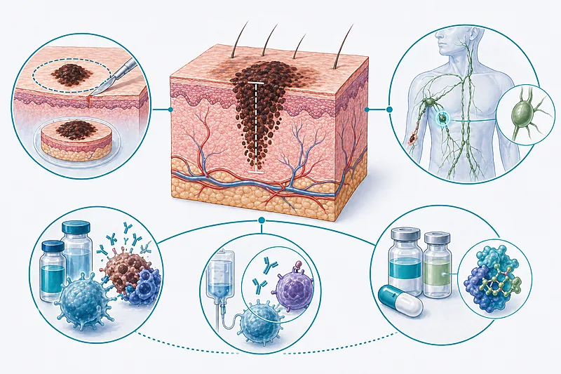
- 1Diagnose every suspicious pigmented lesion with a full-thickness excisional biopsy — shave biopsy underestimates Breslow thickness → Staging
- 2Stage every patient using AJCC 8th edition criteria — Breslow thickness and ulceration remain the dominant prognostic drivers → Staging
- 3Perform wide local excision with margins tied directly to Breslow thickness: 0.5–1 cm for in situ, up to 2 cm for thicker lesions → Wide Local Excision
- 4Discuss sentinel lymph node biopsy in every patient with a primary tumour ≥ 0.8 mm thickness or with high-risk T1b features → Sentinel Node
- 5Do not perform completion lymphadenectomy for a positive sentinel node by default — MSLT-II established active nodal surveillance as the standard → Sentinel Node
- 6Offer adjuvant anti-PD1 immunotherapy to all eligible patients with resected stage IIB/IIC or stage III disease → Adjuvant Therapy
- 7Test for BRAF V600 mutation in all resected stage III disease — dabrafenib plus trametinib is an effective adjuvant alternative in mutation-positive patients → Adjuvant Therapy
- 8Refer every patient with regional or distant disease to a multidisciplinary melanoma board before committing to a treatment sequence → Adjuvant Therapy
- 9Structure follow-up around stage-specific recurrence patterns — most recurrences happen in the first 3 years → Follow-Up
- 10Counsel every patient on rigorous sun protection and skin self-examination — second primary melanoma risk is substantially elevated → Follow-Up
AJCC 8th Staging in Melanoma Treatment
Accurate staging is the backbone of modern melanoma treatment. The AJCC 8th edition, published in 2017, introduced meaningful refinements: T1 was split into T1a and T1b at 0.8 mm rather than 1 mm; mitotic rate was removed from the T category; and stage III substaging was expanded to better reflect survival differences. Getting the stage right at the outset drives margin choice, sentinel node decisions, and adjuvant therapy eligibility.
Perform an excisional biopsy with narrow (1–3 mm) margins for any clinically suspicious pigmented lesion. Shave biopsy frequently transects the lesion base and under-reports Breslow thickness, leading to understaging.
Strong Rec High Evidence NCCN MelanomaStage every confirmed melanoma using AJCC 8th edition criteria. Record Breslow thickness to the nearest 0.1 mm, ulceration status, anatomic location, and satellite/in-transit disease when present.
Strong Rec High Evidence AJCC 8th NCCN MelanomaPerform a full clinical examination, including all regional nodal basins and total body skin, at diagnosis. Palpable lymphadenopathy converts a straightforward wide local excision pathway into one that may require up-front imaging and systemic therapy planning.
Strong Rec Moderate Evidence NCCN MelanomaReserve cross-sectional imaging for stage III and IV disease, symptomatic patients, or before major reconstructive surgery. Routine CT or PET/CT is not indicated for asymptomatic stage I–IIA disease — yield is low and false positives are common.
Moderate Rec Moderate Evidence NCCN MelanomaSend tissue for BRAF V600 mutation testing in every stage III and IV case, and consider it in high-risk stage II disease. Molecular status determines eligibility for targeted therapy and should not be treated as optional.
Strong Rec High Evidence NCCN Melanoma COMBI-AD 2017AJCC 8th T Category at a Glance
| T Category | Breslow Thickness | Ulceration | Surgical Implications |
|---|---|---|---|
| Tis | Melanoma in situ | — | Excision with 0.5–1 cm margin; no SLNB |
| T1a | < 0.8 mm | No | WLE 1 cm; SLNB generally not indicated |
| T1b | < 0.8 mm with ulceration; or 0.8–1.0 mm | Possible | WLE 1 cm; discuss SLNB |
| T2 | > 1.0–2.0 mm | a: no / b: yes | WLE 1–2 cm; SLNB recommended |
| T3 | > 2.0–4.0 mm | a: no / b: yes | WLE 2 cm; SLNB recommended |
| T4 | > 4.0 mm | a: no / b: yes | WLE 2 cm; SLNB recommended; adjuvant discussion earlier |
Wide Local Excision Margins in Melanoma Treatment
Wide local excision (WLE) is the cornerstone of primary surgical melanoma treatment. Modern margin recommendations are narrower than historical wisdom — driven by several randomised trials showing that wider excision does not improve survival, only local control. The aim now is to take enough tissue to prevent local recurrence without over-treating for a benefit that does not exist.
Excise melanoma in situ with a 0.5–1 cm lateral margin. Consider wider margins (up to 1 cm) for lentigo maligna-type lesions on the head and neck where subclinical extension is common.
Strong Rec Moderate Evidence NCCN MelanomaExcise invasive melanomas up to 1.0 mm Breslow thickness with 1 cm margins down to (and including) the deep fascia where anatomically appropriate.
Strong Rec High Evidence NCCN MelanomaUse 1–2 cm margins for melanomas > 1.0–2.0 mm (T2). The specific margin within this range is chosen pragmatically based on anatomical site, closure feasibility, and cosmesis.
Strong Rec High Evidence NCCN MelanomaUse 2 cm margins for melanomas > 2.0 mm (T3 and T4). Wider excision beyond 2 cm does not improve survival and increases reconstruction demands.
Strong Rec High Evidence NCCN MelanomaPlan sentinel node biopsy before or simultaneously with wide local excision when indicated — performing WLE first can alter lymphatic drainage and reduce SLNB accuracy.
Strong Rec Moderate Evidence NCCN MelanomaExcision Margins by Breslow Thickness
| Tumour Thickness | Recommended Margin | Depth | Anatomical Considerations |
|---|---|---|---|
| In situ | 0.5–1 cm | Through dermis | Larger margin for lentigo maligna on face |
| ≤ 1.0 mm | 1 cm | To fascia where practical | Digits & face: individualise; consider functional preservation |
| > 1.0–2.0 mm | 1–2 cm | Include fascia | Narrower end acceptable where cosmesis/function critical |
| > 2.0–4.0 mm | 2 cm | Include fascia | Plan closure/flap before incision; involve plastic surgery |
| > 4.0 mm | 2 cm | Include fascia | Wider not superior; adjuvant discussion from the outset |
Sentinel Node Biopsy and the MSLT-II Legacy
Sentinel lymph node biopsy (SLNB) remains the single most important staging procedure in melanoma. It identifies occult nodal disease, informs prognosis, and now determines eligibility for a growing range of adjuvant systemic therapies. The MSLT-II trial, published in 2017, fundamentally changed what happens next when the sentinel node is positive.
Offer sentinel lymph node biopsy to every patient with a clinically node-negative primary melanoma ≥ 1.0 mm thickness. SLNB is the most accurate method of pathological nodal staging.
Strong Rec High Evidence NCCN Melanoma ASCO/SSODiscuss sentinel node biopsy in patients with T1b primary tumours (0.8–1.0 mm, or < 0.8 mm with ulceration), particularly when high-risk features such as high mitotic rate, lymphovascular invasion, or young age co-exist. The procedure identifies a clinically meaningful subset with occult nodal disease.
Moderate Rec Moderate Evidence NCCN MelanomaDo not routinely perform completion lymphadenectomy after a positive sentinel node. The MSLT-II trial showed no melanoma-specific survival benefit; instead, offer structured nodal-basin ultrasound surveillance and move forward with adjuvant systemic therapy discussions.
Against High Evidence MSLT-II 2017 NCCN MelanomaReserve therapeutic lymph node dissection for patients with clinically or radiologically evident regional nodal disease, or for progression during nodal surveillance after a positive sentinel node.
Strong Rec Moderate Evidence NCCN MelanomaArrange nodal-basin ultrasound surveillance every 4 months for the first 2 years after a positive sentinel node, then every 6 months to year 5. Ultrasound is sensitive, radiation-free, and directly detects the pattern of recurrence most likely to be salvageable.
Moderate Rec Moderate Evidence NCCN MelanomaAdjuvant Systemic Therapy in Melanoma Treatment
Adjuvant systemic therapy has transformed stage II and III melanoma treatment over the past decade. Anti-PD1 immunotherapy (nivolumab, pembrolizumab) and BRAF/MEK-targeted combination therapy (dabrafenib + trametinib for BRAF V600-mutant disease) both produce substantial, durable improvements in recurrence-free survival. The surgeon’s role is to ensure timely referral and shared decision-making with medical oncology.
Refer every eligible patient with resected stage IIB, IIC, or stage III disease for adjuvant anti-PD1 immunotherapy. Pembrolizumab and nivolumab both produce significant improvements in recurrence-free survival over placebo or ipilimumab.
Strong Rec High Evidence KEYNOTE-054 CheckMate 238Offer adjuvant dabrafenib plus trametinib for 12 months as an alternative in resected stage III BRAF V600-mutant disease. The COMBI-AD trial demonstrated durable recurrence-free and overall survival benefit.
Strong Rec High Evidence COMBI-AD 2017Consider neoadjuvant immunotherapy in patients with clinically palpable, resectable stage III disease. Recent randomised data show improved event-free survival when checkpoint inhibitors are given before definitive surgery in this group.
Moderate Rec Moderate Evidence NCCN MelanomaRefer all patients with stage IV metastatic melanoma to a dedicated multidisciplinary melanoma board before any local therapy. First-line systemic options now include dual checkpoint blockade, single-agent anti-PD1, or targeted therapy, with sequencing individualised to disease biology and patient factors.
Strong Rec High Evidence NCCN MelanomaCounsel patients starting immunotherapy explicitly on the spectrum of immune-related adverse events: thyroid dysfunction, colitis, hepatitis, pneumonitis, endocrinopathies, and rare severe reactions. Early recognition and prompt steroid therapy are the pillars of management.
Strong Rec High Evidence NCCN Melanoma ASCO irAEAdjuvant Therapy Options by Stage and Molecular Profile
| Stage | BRAF Status | Preferred Adjuvant | Key Considerations |
|---|---|---|---|
| IIA | Any | Observation usually; trial enrolment if available | Adjuvant therapy not standard; evidence emerging |
| IIB & IIC | Any | Anti-PD1 (pembrolizumab or nivolumab) | KEYNOTE-716 extended approval into stage II |
| III | BRAF wild-type | Anti-PD1 monotherapy | Pembrolizumab or nivolumab; 12 months |
| III | BRAF V600 mutant | Anti-PD1 or dabrafenib + trametinib | Shared decision; pyrexia common with targeted; irAEs with checkpoint |
| IV (resected) | Any | Nivolumab per CheckMate 238 and subsequent data | Highly individualised; MDT decision |
Clinical Decision Pathway
A question-based walk-through from biopsy to adjuvant therapy decision.
Follow-Up and Long-Term Surveillance
Most recurrences in melanoma treatment happen within the first 3 years, but late relapse — even beyond 10 years — is well described. Surveillance intensity should match stage, and follow-up doubles as ongoing skin screening: patients with one primary melanoma are at substantially elevated risk of a second.
| Stage | Years 1–2 | Years 3–5 | Beyond 5 Years | Imaging |
|---|---|---|---|---|
| In situ / IA | 6–12 monthly skin exam | Annually | Annually | Not routine |
| IB / IIA | 3–6 monthly exam + skin | 6–12 monthly | Annually | Consider; per symptoms |
| IIB / IIC / III | 3 monthly exam + nodal basin US | 6 monthly | Annually to year 10 | CT or PET/CT 6–12 monthly in first 2–3 y |
| IV (NED) | Individualised, usually 3 monthly | Individualised | Lifelong specialist follow-up | Ongoing cross-sectional imaging |
Evidence in Context
Key themes from the AJCC 8th edition, MSLT-II, and the landmark adjuvant therapy trials that define modern melanoma treatment.
Why the AJCC 8th Edition Matters
The 8th edition refined T1 substaging at 0.8 mm, removed mitotic rate from T staging (although it remains prognostic), and expanded stage III into four substages (IIIA through IIID) with markedly different survival curves. These refinements are not academic — they directly affect SLNB eligibility, adjuvant therapy eligibility, and follow-up intensity.
MSLT-II: The End of Reflexive Completion Lymphadenectomy
The MSLT-II randomised trial compared immediate completion lymph node dissection with nodal-basin ultrasound surveillance after a positive sentinel node. At three years, melanoma-specific survival was equivalent between the two arms, while complication rates (particularly lymphoedema) were markedly higher in the surgery arm. This result reshaped the surgical management of sentinel-node-positive disease almost overnight.
Adjuvant Immunotherapy: CheckMate 238 and KEYNOTE-054
CheckMate 238 compared adjuvant nivolumab with ipilimumab in resected stage III/IV disease and showed that nivolumab produced substantially better recurrence-free survival with less toxicity. KEYNOTE-054 compared adjuvant pembrolizumab with placebo in resected stage III and showed durable recurrence-free survival benefit. Together these studies established anti-PD1 monotherapy as the standard adjuvant backbone.
Adjuvant Targeted Therapy: COMBI-AD
The COMBI-AD trial established adjuvant dabrafenib plus trametinib for 12 months as an effective option in resected stage III BRAF V600-mutant disease, with durable recurrence-free and overall survival benefit. The choice between anti-PD1 and BRAF/MEK targeted therapy in BRAF-mutant patients remains a shared decision, weighing long-term efficacy, toxicity profiles, and patient preference.
What We Still Don’t Know
The optimal sequence of neoadjuvant versus adjuvant immunotherapy is an active area of research. Long-term outcomes beyond 10 years of checkpoint inhibitor therapy are still accumulating. The best strategy for stage IIA disease (where adjuvant therapy is not standard but recurrence risk is non-trivial) remains debated, and biomarkers for predicting response or immune-related toxicity are not yet clinically actionable at the individual patient level.
References
- 1.Gershenwald JE, Scolyer RA, Hess KR, et al. Melanoma staging: Evidence-based changes in the American Joint Committee on Cancer eighth edition cancer staging manual. CA Cancer J Clin. 2017;67(6):472–492. doi:10.3322/caac.21409
- 2.Faries MB, Thompson JF, Cochran AJ, et al. Completion Dissection or Observation for Sentinel-Node Metastasis in Melanoma. N Engl J Med. 2017;376(23):2211–2222. doi:10.1056/NEJMoa1613210
- 3.Weber J, Mandala M, Del Vecchio M, et al. Adjuvant Nivolumab versus Ipilimumab in Resected Stage III or IV Melanoma. N Engl J Med. 2017;377(19):1824–1835. doi:10.1056/NEJMoa1709030
- 4.Eggermont AMM, Blank CU, Mandala M, et al. Adjuvant Pembrolizumab versus Placebo in Resected Stage III Melanoma. N Engl J Med. 2018;378(19):1789–1801. doi:10.1056/NEJMoa1802357
- 5.Long GV, Hauschild A, Santinami M, et al. Adjuvant Dabrafenib plus Trametinib in Stage III BRAF-Mutated Melanoma. N Engl J Med. 2017;377(19):1813–1823. doi:10.1056/NEJMoa1708539
- 6.Luke JJ, Rutkowski P, Queirolo P, et al. Pembrolizumab versus placebo as adjuvant therapy in resected stage IIB or IIC melanoma (KEYNOTE-716): a randomised, double-blind, phase 3 trial. Lancet. 2022;399(10336):1718–1729. doi:10.1016/S0140-6736(22)00562-1
How to Read the Evidence Tags
Every recommendation in this Practice Update carries three inline tags: recommendation strength, evidence quality, and source. These are Medaptly’s own simplified interpretations — not reproductions of any single guideline body’s classification system.
Recommendation Strength
| Tag | What It Means |
|---|---|
| Strong Rec | High-quality evidence broadly supports this action. |
| Moderate Rec | The weight of evidence favours this action. |
| Conditional Rec | The benefit is less certain — individualise to the patient. |
| Against | Evidence shows no benefit or potential harm. |
Evidence Quality
| Tag | What It Means |
|---|---|
| High Evidence | Multiple well-designed RCTs or high-quality meta-analyses. |
| Moderate Evidence | Single RCT or large observational studies. |
| Low Evidence | Expert consensus or small studies. |