
Carotid Stenosis Management: 8 Essential Clinical Decisions
Clinical Practice Update — Stratifying Risk and Choosing Revascularization
This is an original clinical education article informed by current guidelines and evidence. See References below for source documents.
- Clinical Focus
- Evidence-based carotid stenosis management in symptomatic and asymptomatic adults
- Target Audience
- Neurologists, stroke physicians, vascular surgeons, primary care physicians, residents
- Setting
- Outpatient neurology, stroke unit, vascular clinic, primary care
- Source Evidence
- •AHA/ASA Guidelines for the Prevention of Stroke in Patients With Stroke and TIA (2021)
- •Society for Vascular Surgery (SVS) Guidelines on Extracranial Cerebrovascular Disease (2022)
- •ESVS Clinical Practice Guidelines on Atherosclerotic Carotid and Vertebral Artery Disease (2023)
- •ACST-2 Trial — Stenting vs Endarterectomy for Asymptomatic Carotid Stenosis (Lancet, 2021)
- •CREST 10-Year Outcomes (NEJM, 2016)
Key Clinical Takeaways
Effective carotid stenosis management in 2026 rests on three intertwined questions: is the lesion symptomatic, how severe is it, and does the patient stand to gain more from revascularization than from optimised medical therapy alone? The points below distil current evidence into actionable bedside rules.
- 1Define symptom status first — the entire decision tree turns on whether the patient has had an ipsilateral event in the last 6 months → Risk Stratification
- 2Confirm any duplex finding above 70% with CTA or MRA before booking a procedure → Risk Stratification
- 3High-intensity statin therapy belongs in every patient with significant carotid disease, regardless of baseline LDL-C → Medical Therapy
- 4Revascularize symptomatic 70–99% stenosis within 14 days — benefit decays sharply with delay → Symptomatic Carotid Stenosis
- 5For asymptomatic disease, the bar for intervention is much higher — favour medical therapy unless ipsilateral risk markers are present → Asymptomatic Carotid Stenosis
- 6Prefer CEA over CAS in patients above 70 years with surgically accessible anatomy → CEA vs CAS
- 7Consider CAS or TCAR when the neck is hostile, the patient is high surgical risk, or restenosis follows prior CEA → CEA vs CAS
- 8Do not screen the general population — the harms of incidental asymptomatic disease detection outweigh the benefits → Asymptomatic Carotid Stenosis
Risk Stratification in Carotid Stenosis Management
Every clinical decision in carotid stenosis management starts with two binary classifications: symptomatic versus asymptomatic, and stenosis severity. Symptom status is the dominant variable — a 70% lesion behaves very differently before and after the first ischemic event. A patient with a symptomatic 70–99% stenosis carries a roughly one-in-four risk of recurrent stroke within two years on medical therapy alone; the same anatomy in an asymptomatic patient carries a stroke risk closer to 1% per year under current medical regimens.
Defining “symptomatic” is more nuanced than it sounds. The classic window is any ipsilateral hemispheric stroke, transient ischemic attack, or retinal ischemic event in the preceding 6 months. Vague non-focal symptoms (presyncope, isolated dizziness, “fuzzy” episodes) should not trigger this reclassification — doing so inflates the perceived stroke risk and pushes patients toward unnecessary procedures.
Reclassify a patient as symptomatic only if they have had an ipsilateral hemispheric stroke, TIA, or retinal ischemic event (including amaurosis fugax) within the past 6 months. Non-focal symptoms do not qualify.
Strong Rec High Evidence AHA/ASA 2021Use carotid duplex ultrasound as the first-line imaging modality, then confirm any ≥70% finding with CT angiography or contrast MR angiography before scheduling revascularization.
Strong Rec High Evidence SVS 2022Quantify stenosis using NASCET-style criteria (distal lumen as reference). Convert ECST measurements when reviewing older studies: an ECST 70% lesion roughly corresponds to a NASCET 50% lesion.
Strong Rec High Evidence ESVS 2023Document plaque features that modify risk — ulceration, intraplaque haemorrhage on MR, and contralateral occlusion all raise the ipsilateral stroke risk and may shift an asymptomatic patient toward intervention.
Moderate Rec Moderate Evidence SVS 2022Medical Therapy: The Foundation of Carotid Stenosis Management
Intensive medical therapy is not the alternative to revascularization — it is the foundation under it. The dramatic drop in stroke rates among asymptomatic patients over the past two decades is largely attributable to better statins, antiplatelets, and blood pressure control. Whatever else carotid stenosis management requires, it requires this first.
The principles overlap heavily with secondary stroke prevention more broadly: lipid lowering, platelet inhibition, blood pressure control, glycemic management, and tobacco cessation. Where carotid stenosis management diverges is in the intensity of the LDL-C target and the role of short-course dual antiplatelet therapy after a minor symptomatic event.
Start high-intensity statin therapy (atorvastatin 40–80 mg or rosuvastatin 20–40 mg daily) for every adult with significant carotid stenosis, regardless of baseline LDL-C.
Strong Rec High Evidence AHA/ASA 2021Target LDL-C below 70 mg/dL (1.8 mmol/L) for carotid stenosis management overall, and below 55 mg/dL (1.4 mmol/L) after an ischemic stroke or TIA. Add ezetimibe or a PCSK9 inhibitor if the target is not reached.
Strong Rec High Evidence ESVS 2023Prescribe single antiplatelet therapy — aspirin 81 mg daily or clopidogrel 75 mg daily — for every patient with carotid stenosis. The two agents are broadly equivalent for long-term use.
Strong Rec High Evidence AHA/ASA 2021Initiate dual antiplatelet therapy (aspirin plus clopidogrel) for 21–30 days after a high-risk TIA (ABCD2 ≥4) or minor non-cardioembolic stroke (NIHSS ≤3), then return to monotherapy.
Strong Rec High Evidence AHA/ASA 2021Avoid extending dual antiplatelet therapy beyond 90 days for indications other than recent stenting — bleeding risk rises sharply without proportional benefit.
Against Moderate Evidence AHA/ASA 2021Target blood pressure below 130/80 mmHg as part of long-term carotid stenosis management. Hold off on aggressive acute lowering in the first 24–48 hours after an ischemic event unless BP exceeds 220/120 mmHg.
Strong Rec Moderate Evidence AHA/ASA 2021Counsel every patient with carotid stenosis on tobacco cessation at every visit. Smoking accelerates plaque progression more than any other modifiable factor.
Strong Rec High Evidence AHA/ASA 2021Symptomatic Carotid Stenosis: Revascularization Thresholds
The data for symptomatic disease are among the most robust in vascular neurology. NASCET and ECST established — and decades of subsequent work have reinforced — that revascularization sharply reduces recurrent stroke when stenosis is severe and the patient has had a recent ipsilateral event. The window matters: the absolute benefit of intervention is highest within 14 days of the index event and falls steeply after that.
In practice this means that the TIA workup, imaging, and surgical referral should run in parallel, not in series. A patient who presents on Monday with a TIA and an ipsilateral 80% stenosis should ideally be on the operating table or in the cath lab by the following week — not waiting six weeks for a routine vascular surgery slot.
Refer for revascularization within 14 days of the index ipsilateral event in patients with 70–99% symptomatic carotid stenosis and a life expectancy beyond 1 year.
Strong Rec High Evidence AHA/ASA 2021 ESVS 2023Consider revascularization in symptomatic 50–69% stenosis, weighing age, sex, plaque morphology, and perioperative risk. Men derive more benefit than women in this severity band; older patients tend to benefit more from CEA than CAS.
Moderate Rec Moderate Evidence NASCETDo not revascularize symptomatic stenosis below 50% — medical therapy alone is preferred. The procedural risk outweighs any reduction in recurrent stroke at this severity.
Against High Evidence NASCETDelay revascularization beyond 48 hours after large completed strokes with significant penumbra or haemorrhagic transformation — reperfusion of fragile tissue can precipitate hyperperfusion syndrome and intracerebral haemorrhage.
Moderate Rec Moderate Evidence SVS 2022Asymptomatic Carotid Stenosis: Who Truly Benefits
The harder question in carotid stenosis management is what to do with the asymptomatic lesion. ACAS and ACST showed roughly a halving of stroke risk with CEA in selected patients on the medical regimens of the 1990s, but contemporary medical therapy has narrowed that gap substantially. Many patients with 70–99% asymptomatic stenosis treated with modern intensive medical therapy now have annual ipsilateral stroke rates around 1%, leaving little room for a procedural intervention to add net benefit.
The clinical judgment, then, is whether your individual patient resembles the trial cohorts in whom revascularization helped, or whether they are a patient for whom medical therapy alone is the better choice. Three pieces of information drive this judgment: life expectancy, plaque biology, and the local perioperative complication rate.
Reserve revascularization for asymptomatic stenosis ≥70% only when life expectancy exceeds 5 years AND local periprocedural stroke/death rate is documented below 3%.
Moderate Rec Moderate Evidence AHA/ASA 2021 SVS 2022Manage asymptomatic stenosis below 70% with intensive medical therapy alone in the great majority of patients. Revascularization at lower severities adds risk without consistent benefit.
Strong Rec Moderate Evidence ACST-2Consider revascularization at lower asymptomatic stenosis severities when high-risk markers cluster — intraplaque haemorrhage on MR, silent ipsilateral infarcts on imaging, microemboli on transcranial Doppler, or rapid progression on serial duplex.
Conditional Rec Low Evidence SVS 2022Do not screen the general adult population for asymptomatic carotid stenosis. The false-positive rate generates more harm from unnecessary downstream procedures than benefit from earlier detection.
Against High Evidence USPSTF 2021CEA vs CAS: Choosing the Right Procedure
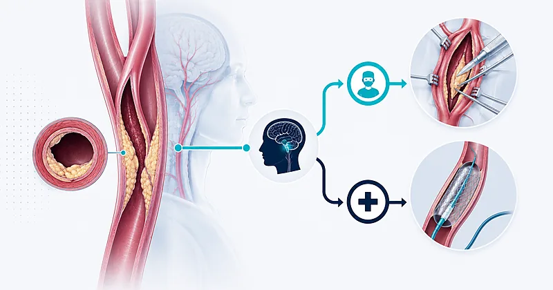
Once revascularization is on the table, the choice between carotid endarterectomy (CEA) and carotid artery stenting (CAS) hinges chiefly on age and anatomy. The headline from CREST and its 10-year follow-up is that the long-term composite of stroke, MI, and death is similar between procedures, but the trade-offs in periprocedural risk differ in important ways: CAS carries a higher periprocedural stroke risk, CEA carries a higher periprocedural MI risk.
Age is the most consistent effect modifier. Patients above roughly 70 years have a higher periprocedural stroke risk with CAS than with CEA — thought to reflect more friable, calcified plaques and tortuous arches. Below 70, the two procedures perform similarly in terms of stroke. Anatomy modifies the calculus further: a hostile neck (prior radiation, tracheostomy, contralateral laryngeal nerve palsy) favours an endovascular approach; a tortuous aortic arch or heavy arch calcification favours an open one.
Transcarotid artery revascularization (TCAR) has emerged as a third option for carotid stenosis management in patients at high surgical risk. It combines a small cervical incision with flow reversal during stenting, and registry data suggest periprocedural stroke rates comparable to CEA in selected patients.
Prefer CEA over CAS in patients above 70 years with surgically accessible carotid anatomy. CAS in this age group carries roughly twice the periprocedural stroke risk of CEA.
Strong Rec High Evidence CRESTConsider CAS or TCAR in patients with a hostile neck (prior neck radiation, prior CEA with restenosis, contralateral laryngeal nerve palsy, surgically inaccessible high carotid bifurcation) or those at very high surgical/cardiac risk.
Moderate Rec Moderate Evidence SVS 2022Perform CEA or CAS only in centres with documented periprocedural stroke/death rates below 3% for asymptomatic disease and below 6% for symptomatic disease. Audit local outcomes annually.
Strong Rec Moderate Evidence AHA/ASA 2021Consider TCAR as an alternative to transfemoral CAS in patients at high surgical risk for CEA — registry data show periprocedural stroke rates comparable to open repair in selected patients.
Moderate Rec Moderate Evidence SVS 2022Continue aspirin throughout the perioperative period for CEA and CAS — do not hold antiplatelet therapy for any carotid procedure.
Strong Rec High Evidence AHA/ASA 2021Add clopidogrel for at least 30 days after stenting in addition to aspirin, then return to single antiplatelet therapy. Tight blood pressure targets after stroke and stenting reduce hyperperfusion injury — aim for systolic 120–140 mmHg in the first week post-procedure.
Strong Rec Moderate Evidence CRESTCEA vs CAS: Patient-Factor Matrix
| Patient Factor | Favours CEA | Favours CAS or TCAR | Practical Note |
|---|---|---|---|
| Age >70 years | Yes — significantly lower periprocedural stroke | No, unless surgically prohibitive | The single strongest discriminator in CREST |
| Prior neck radiation | No — scarred fascial planes | Yes | TCAR often preferred over transfemoral CAS |
| Restenosis after prior CEA | No — reoperation difficult | Yes | Confirm true restenosis vs new contralateral disease |
| High bifurcation (above C2) | No | Yes | Mandibular subluxation rarely worth it |
| Severe cardiac comorbidity | No — CEA has higher periprocedural MI risk | Yes, especially TCAR under local anaesthesia | Optimise cardiology evaluation pre-procedure |
| Heavy aortic arch calcification | Yes | No (transfemoral CAS); TCAR bypasses the arch | TCAR mitigates arch-related stroke risk |
| Symptomatic disease, age <70 | Yes — default | Acceptable when CEA contraindicated | CREST showed equivalent long-term outcomes |
Clinical Decision Pathway for Carotid Stenosis Management
A practical, question-based pathway for carotid stenosis management. Walk through the questions in order; do not skip ahead.
Thresholds at a Glance
The summary table below crosses symptom status with stenosis severity and shows what most contemporary guidelines recommend — with the practical caveats that distinguish carotid stenosis management at the bedside from what reads cleanly on paper.
| Patient Group | Stenosis Severity | First-Line Approach | Time to Procedure | Key Caveats |
|---|---|---|---|---|
| Symptomatic | 70–99% | Revascularize + intensive medical therapy | Within 14 days | Avoid near-occlusion (string sign); revascularization rarely helps |
| Symptomatic | 50–69% | Individualised — revascularize if features favour benefit | Within 14 days if proceeding | Men, age 65–75, recent stroke > TIA all favour intervention |
| Symptomatic | <50% | Medical therapy alone | N/A | Reassess if recurrent ipsilateral events on optimal therapy |
| Asymptomatic | 70–99% | Medical therapy; selectively revascularize | Elective (4–8 weeks) | Requires >5-year life expectancy AND audited <3% complication rate |
| Asymptomatic | 60–69% | Medical therapy alone | N/A | Reconsider only with multiple high-risk plaque features |
| Asymptomatic | <60% | Medical therapy alone | N/A | Annual surveillance duplex only if progression suspected |
- “Symptomatic” requires an ipsilateral event within the last 6 months.
- All percentages refer to NASCET-style measurements on confirmatory CTA/MRA.
- “Medical therapy” throughout means high-intensity statin, single antiplatelet (with short-course DAPT after qualifying events), BP control to <130/80, tobacco cessation, and glycemic optimisation.
Monitoring and Follow-Up
Carotid stenosis management does not end with the operation or with starting the statin. Post-revascularization surveillance and long-term medical optimisation determine whether the early procedural risk paid off.
Schedule surveillance duplex ultrasound at 1, 6, and 12 months after revascularization, then annually for at least 5 years. Re-image sooner if neurological symptoms recur.
Strong Rec Low Evidence SVS 2022Continue lifelong high-intensity statin and single antiplatelet therapy after revascularization — the goal of carotid stenosis management is to slow systemic atherosclerosis, not just to repair one anatomical lesion.
Strong Rec High Evidence AHA/ASA 2021| Parameter | When to Check | Action Threshold | Common Pitfalls |
|---|---|---|---|
| Duplex ultrasound | 1, 6, 12 months post-procedure; then annually | Restenosis >70% or rapid progression → CTA confirmation | Early post-op duplex can overestimate stenosis from edema |
| LDL-C | 4–12 weeks after starting/changing statin; then every 6–12 months | >70 mg/dL (or >55 post-stroke) → intensify therapy | Don’t wait for LDL goals to “settle” — add ezetimibe early if behind target |
| Blood pressure | Every visit; home BP log | >130/80 mmHg → uptitrate or add agent | Watch for orthostasis — common in older patients with bilateral disease |
| Neurological symptoms | Every visit; patient-initiated for new events | Any new ipsilateral focal symptoms → urgent imaging | Educate patients explicitly — many don’t recognise amaurosis fugax as significant |
| Tobacco status | Every visit | Active smoking → offer NRT, varenicline, or bupropion | Most patients understate use — ask specifically about vaping |
Evidence in Context
Where the major guidelines agree, where they diverge, and what the trial data actually show for carotid stenosis management in 2026.
Where AHA/ASA, SVS, and ESVS agree
All three contemporary frameworks agree on the broad architecture of carotid stenosis management: symptom status drives urgency, severity drives the procedural threshold, optimised medical therapy underpins both arms, and centre quality (audited periprocedural complication rate) is a prerequisite for offering revascularization.
They also agree on the symptomatic threshold — 70–99% severe stenosis warrants revascularization within 14 days, and intervention should be considered down to 50% with patient-specific factors weighing in.
Where the guidelines diverge
Asymptomatic threshold: ESVS 2023 leans more conservatively, restricting intervention to asymptomatic patients with one or more “high-risk for stroke on medical therapy” features. AHA/ASA 2021 is somewhat more permissive, allowing revascularization in selected asymptomatic 70–99% lesions on the basis of life expectancy and centre quality alone.
LDL-C target: ESVS 2023 endorses the <55 mg/dL target after ischemic stroke; AHA/ASA 2021 emphasises <70 mg/dL as the general threshold with intensification toward 55 in higher-risk patients.
CAS in asymptomatic patients: SVS 2022 is more open to CAS as an alternative to CEA than ESVS 2023, which restricts CAS in asymptomatic disease largely to centres with proven low complication rates and patients at high surgical risk.
What CREST and its 10-year follow-up actually showed
CREST randomised over 2,500 symptomatic and asymptomatic patients to CEA or CAS. The primary composite endpoint (periprocedural stroke, MI, death; ipsilateral stroke through 4 years) showed no overall difference between the two procedures. But the trade-off pattern was clear: periprocedural stroke favoured CEA, periprocedural MI favoured CAS.
The 10-year follow-up confirmed that this near-equivalence held over time, with no late divergence between procedures — meaning that once a patient survives the periprocedural period, long-term durability is broadly similar.
Why ACST-2 didn’t end the asymptomatic debate
ACST-2 randomised over 3,600 asymptomatic patients between CEA and CAS — not between revascularization and medical therapy. It confirmed that the two procedures yield similar non-disabling stroke and death rates at 5 years but did not address whether either is better than modern intensive medical therapy alone. That question is the subject of ongoing trials (notably CREST-2), which are likely to reshape recommendations on asymptomatic disease over the next several years.
The case against general population screening
The USPSTF in 2021 reaffirmed its recommendation against screening asymptomatic adults for carotid stenosis. The reasoning is straightforward: prevalence of severe asymptomatic stenosis is low in the general population, ultrasound has imperfect specificity, and the cascade of confirmatory imaging, anxiety, and downstream procedures has documented harms. Screening makes sense only when a patient has multiple vascular risk factors and the result will plausibly change management.
References
- 1.Kleindorfer DO, Towfighi A, Chaturvedi S, et al. 2021 Guideline for the Prevention of Stroke in Patients With Stroke and Transient Ischemic Attack: A Guideline From the American Heart Association/American Stroke Association. Stroke. 2021;52(7):e364–e467. doi:10.1161/STR.0000000000000375
- 2.AbuRahma AF, Avgerinos ED, Chang RW, et al. Society for Vascular Surgery clinical practice guidelines for management of extracranial cerebrovascular disease. J Vasc Surg. 2022;75(1S):4S–22S. doi:10.1016/j.jvs.2021.04.073
- 3.Naylor AR, Rantner B, Ancetti S, et al. Editor’s Choice — European Society for Vascular Surgery (ESVS) 2023 Clinical Practice Guidelines on the Management of Atherosclerotic Carotid and Vertebral Artery Disease. Eur J Vasc Endovasc Surg. 2023;65(1):7–111. doi:10.1016/j.ejvs.2022.04.011
- 4.Halliday A, Bulbulia R, Bonati LH, et al. Second asymptomatic carotid surgery trial (ACST-2): a randomised comparison of carotid artery stenting versus carotid endarterectomy. Lancet. 2021;398(10305):1065–1073. doi:10.1016/S0140-6736(21)01910-3
- 5.Brott TG, Howard G, Roubin GS, et al. Long-Term Results of Stenting versus Endarterectomy for Carotid-Artery Stenosis. N Engl J Med. 2016;374(11):1021–1031. doi:10.1056/NEJMoa1505215
- 6.US Preventive Services Task Force. Screening for Asymptomatic Carotid Artery Stenosis: US Preventive Services Task Force Recommendation Statement. JAMA. 2021;325(5):476–481. doi:10.1001/jama.2020.26988
How to Read the Evidence Tags
Every recommendation carries three tags — recommendation strength, evidence quality, and source — using Medaptly’s own simplified interpretation of the underlying classification systems.
Recommendation Strength
| Tag | What It Means |
|---|---|
| Strong Rec | High-quality evidence broadly supports this action across most patients. |
| Moderate Rec | The weight of evidence favours this action; individualisation reasonable. |
| Conditional Rec | Benefit is less certain — weigh patient-specific factors. |
| Against | Evidence shows no benefit or net harm. |
Evidence Quality
| Tag | What It Means |
|---|---|
| High Evidence | Multiple well-designed RCTs or high-quality meta-analyses. |
| Moderate Evidence | Single RCT or large observational studies. |
| Low Evidence | Expert consensus or small studies. |