
Shoulder Dystocia: Stepwise Maneuvers and Team Response
Clinical Practice Update — Recognition, Sequential Delivery Maneuvers, and Coordinated Team Response
This is an original clinical education article informed by current guidelines and evidence. See References below for source documents.
- Clinical Focus
- Recognition and stepwise shoulder dystocia maneuvers in the immediate intrapartum period
- Target Audience
- Obstetricians, midwives, family physicians, emergency physicians, labor-ward nurses, residents
- Setting
- Labor and delivery units, birth centers, emergency departments
- Source Evidence
- •ACOG Practice Bulletin No. 178 — Shoulder Dystocia (2017)
- •RCOG Green-top Guideline No. 42 — Shoulder Dystocia (2012)
- •Grobman et al. Critical Evaluation of Maneuvers — AJOG (2024)
- •FEBRASGO Position Statement — Management of Shoulder Dystocia (2022)
Key Clinical Takeaways
Effective use of shoulder dystocia maneuvers depends on three things happening almost at once: instant recognition, a calm escalation through a rehearsed sequence, and a coordinated team. Because the condition is largely unpredictable, every clinician attending births should be able to move through the steps below from memory, under pressure, with the clock running.
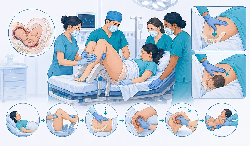
- 1Recognize the dystocia at the moment gentle downward traction fails to deliver the anterior shoulder — do not wait for the next contraction to “try again.”
- 2Call for help out loud, name the emergency, and start the clock the instant the shoulder is recognized as stuck.
- 3Perform the McRoberts maneuver first — it is simple, fast, and resolves a large share of cases.
- 4Add suprapubic pressure directed downward and laterally to disimpact the anterior shoulder.
- 5Move to internal shoulder dystocia maneuvers — delivery of the posterior arm and internal rotation — when external steps fail.
- 6Never apply fundal pressure and avoid forceful lateral traction on the fetal head — both worsen impaction and risk brachial plexus injury.
- 7Document the head-to-body interval, the order and timing of each maneuver, and which shoulder was anterior.
- 8Build team fluency through regular simulation training, which improves both clinical outcomes and the quality of documentation.
Recognition: When Shoulder Dystocia Maneuvers Become Necessary
Recognition is the trigger for everything that follows. Shoulder dystocia is best understood as a bony impaction: the anterior shoulder lodges behind the pubic symphysis, or less often the posterior shoulder catches on the sacral promontory, so the trunk cannot follow the head. The diagnosis is clinical and is made the moment routine gentle traction fails.
Diagnose shoulder dystocia when gentle downward traction fails to deliver the anterior shoulder after the head has emerged, or when the head retracts tightly against the perineum (the turtle sign). Treat either finding as confirmation rather than waiting for further attempts.
Strong Rec Low Evidence ACOG 2017 RCOG 2012Anticipate, but do not rely on, known associations such as fetal macrosomia, prior shoulder dystocia, diabetes, and prolonged second stage. Their predictive value is poor, so preparedness at every birth matters more than risk-scoring any single labor.
Moderate Rec Moderate Evidence ACOG 2017Announce the diagnosis explicitly and call for help the moment dystocia is recognized. State the problem in plain words so the whole room shifts into the emergency sequence together.
Strong Rec Moderate Evidence RCOG 2012First-Line Shoulder Dystocia Maneuvers
The opening shoulder dystocia maneuvers are external, non-invasive, and resolve most cases. They share a logic: change the geometry of the pelvis and shoulder girdle before reaching inside. Apply them in a deliberate order, giving each roughly thirty seconds before escalating.
Perform the McRoberts maneuver first: hyperflex and abduct the maternal hips so the thighs come toward the abdomen. This rotates the symphysis cephalad and straightens the sacrum, frequently freeing the anterior shoulder on its own.
Strong Rec Moderate Evidence ACOG 2017 RCOG 2012Apply suprapubic pressure with McRoberts in place. An assistant presses just above the symphysis, aiming the force downward and toward the side of the fetal back at roughly a 45-degree angle, to adduct and disimpact the anterior shoulder.
Strong Rec Moderate Evidence ACOG 2017Consider an episiotomy only when more room is needed for the operator’s hand to perform internal maneuvers. It does not relieve a bony impaction by itself, so reserve it for access rather than performing it routinely.
Conditional Rec Low Evidence RCOG 2012Do not apply fundal pressure at any point. It drives the impacted shoulder harder against the symphysis, raises the risk of uterine rupture, and has no role in resolving the dystocia.
Against Moderate Evidence ACOG 2017 RCOG 2012Internal Rotation and Posterior Arm Delivery
When external steps do not free the shoulder within about a minute, escalate to internal maneuvers. These act inside the pelvis to either reduce the shoulder-to-shoulder diameter or remove one arm entirely. There is no single mandated order; choose what the room and your hands allow, and switch promptly if one approach stalls.
Deliver the posterior arm: insert a hand along the fetal back, locate the posterior forearm, flex it across the chest, and sweep it out. Removing one arm narrows the impacted girdle substantially and resolves a high proportion of otherwise refractory cases.
Strong Rec Moderate Evidence ACOG 2017Perform internal rotation by pressing on the back or front of a fetal shoulder to turn the girdle into the wider oblique diameter of the pelvis. Pressure behind the posterior shoulder (Woods screw) or behind the anterior shoulder (Rubin) both work; pick whichever surface your fingers reach first.
Moderate Rec Moderate Evidence ACOG 2017 AJOG 2024Combine rotation with posterior arm delivery when either alone is incomplete. Rotating the trunk a few degrees often converts an unreachable posterior arm into one you can flex and sweep.
Moderate Rec Low Evidence AJOG 2024Move the patient onto all fours (the Gaskin position) if internal access is difficult, particularly in a non-anesthetized patient. The change in pelvic orientation can itself dislodge the shoulder and improves the operator’s reach to the posterior arm.
Conditional Rec Low Evidence RCOG 2012 FEBRASGO 2022When First-Line Steps Fail: Rescue Options
A small number of cases resist every standard maneuver. The options below carry real maternal and fetal morbidity and are reserved for genuine impasse, ideally with senior obstetric, anesthetic, and neonatal teams present.
Consider deliberate fracture of the fetal clavicle to reduce the shoulder girdle when standard maneuvers have failed. Direct pressure outward, away from the lung, to limit the risk of pneumothorax.
Conditional Rec Low Evidence FEBRASGO 2022Reserve the Zavanelli maneuver (cephalic replacement followed by cesarean) and symphysiotomy for catastrophic, otherwise undeliverable dystocia. Both carry high rates of maternal morbidity, including severe perineal lacerations and urinary tract injury, and demand an explicit risk-benefit judgment.
Conditional Rec Low Evidence RCOG 2012 FEBRASGO 2022Clinical Decision Pathway
A question-based way to move through shoulder dystocia maneuvers under time pressure. Work through the questions in order, escalating roughly every thirty seconds.
Team Response and Role Assignment
Shoulder dystocia is a team event. Outcomes improve when each person knows their job before the emergency begins. The table below frames responsibilities by role rather than by maneuver, so a glance tells each team member where to stand and what to do.
| Role | Primary Task | Where to Stand | Watch Out For |
|---|---|---|---|
| Lead accoucheur | Calls the sequence, performs internal maneuvers | At the perineum | Avoid excessive head traction |
| Assistant 1 | McRoberts leg flexion | At one maternal leg | Full hyperflexion, not just abduction |
| Assistant 2 | Suprapubic pressure | At the maternal flank, facing the back | Never confuse with fundal pressure |
| Scribe | Timekeeping and contemporaneous notes | At the bedside, visible clock | Record each maneuver as it happens |
| Neonatal team | Prepare resuscitation, assess the arm | At the warmer | Examine for brachial plexus deficit |
Maneuver Reference at a Glance
A consolidated reference organized by the mechanism each maneuver uses, with a practical execution note and the most common pitfall for each. This is a memory aid for drills, not a rigid order of operations.
| Maneuver | How It Works | Execution Note | Common Pitfall |
|---|---|---|---|
| McRoberts | Rotates symphysis cephalad, flattens sacrum | Thighs sharply onto the abdomen | Abducting without true hyperflexion |
| Suprapubic pressure | Adducts and disimpacts anterior shoulder | Push down and toward fetal back | Pressing midline or too high |
| Posterior arm delivery | Removes an arm, narrows the girdle | Flex at the elbow, sweep across chest | Pulling the hand and fracturing the forearm |
| Internal rotation | Turns girdle into wider oblique diameter | Pressure on a shoulder, front or back | Pushing on the head instead of the shoulder |
| All fours (Gaskin) | Reorients pelvis, opens sacral space | Hands and knees, deliver posterior shoulder | Delaying the switch when access is poor |
Immediate Aftercare and Documentation
The minutes after delivery shape both clinical safety and medicolegal defensibility. Maternal and neonatal complications must be sought actively, and the record must be written while memory is fresh.
| What to Assess | When | Why It Matters | Common Pitfall |
|---|---|---|---|
| Maternal genital tract | Immediately postpartum | Higher risk of postpartum hemorrhage and third/fourth-degree tears | Assuming an easy repair means no injury |
| Neonatal arm and clavicle | At delivery and on newborn exam | Detects brachial plexus injury and fracture early | Missing a subtle reduced-movement arm |
| Cord gases | At delivery when feasible | Objective record of acid-base status | Forgetting to sample in the rush |
| Structured event record | As soon as the patient is stable | Captures sequence, timing, and personnel | Writing it hours later from memory |
Document contemporaneously: record the head-to-body interval, which shoulder was anterior, the order and timing of each maneuver, who performed it, and the neonatal condition at birth. A structured proforma improves completeness.
Strong Rec Moderate Evidence ACOG 2017 RCOG 2012Debrief the team and the family after the event. Explain what happened, examine the newborn for any deficit, and arrange follow-up for an arm that is not moving normally.
Moderate Rec Low Evidence RCOG 2012Evidence in Context
What the evidence supports, where the major guidance agrees, and where genuine uncertainty remains.
Where ACOG and RCOG Agree
Both bodies converge on the essentials: shoulder dystocia is unpredictable and largely unpreventable, McRoberts is the sensible opening maneuver, fundal pressure is prohibited, and structured documentation plus simulation training improve outcomes. The shared message is that fluent sequencing, not prediction, protects mothers and babies.
Is There a Single Best Second Maneuver?
Older guidance held that no maneuver is clearly superior after McRoberts. More recent analysis argues that delivery of the posterior arm has among the highest success rates and that direct internal rotation may require less traction force than repeated external attempts. No randomized trials settle the question, so the practical answer is to use whichever internal maneuver your hands can reach and to switch quickly if it stalls.
How Strong Is the Evidence Base?
Most of what guides shoulder dystocia management comes from observational series, registry data, and biomechanical reasoning rather than randomized comparisons. This is why so many recommendations carry a low or moderate evidence tag despite being strongly recommended: the clinical logic and consensus are firm even where trial data are thin.
What Does Simulation Training Actually Change?
Multi-professional drills on high-fidelity mannequins have been associated with faster, more correct maneuver use, better team communication, and improved documentation. Several programs report reductions in transient brachial plexus injury after introducing structured training, which is why both guidelines actively recommend it.
References
- 1.American College of Obstetricians and Gynecologists. Shoulder dystocia. Practice Bulletin No. 178. Obstet Gynecol. 2017;129(5):e123–e133. doi:10.1097/AOG.0000000000002043
- 2.Royal College of Obstetricians and Gynaecologists. Shoulder dystocia. Green-top Guideline No. 42. 2nd ed. London: RCOG; 2012. rcog.org.uk/media/ewgpnmio/gtg_42.pdf
- 3.Grobman WA, Bjorklund AB. A critical evaluation of the external and internal maneuvers for resolution of shoulder dystocia. Am J Obstet Gynecol. 2024;230(3S):S1011–S1018. doi:10.1016/j.ajog.2022.07.024
- 4.Nomura RMY, et al. Management of shoulder dystocia. FEBRASGO Position Statement. Rev Bras Ginecol Obstet. 2022;44(7):723–736. doi:10.1055/s-0042-1755458
- 5.Hill MG, Cohen WR. Shoulder dystocia: prediction and management. Womens Health (Lond). 2016;12(2):251–261. doi:10.2217/whe.15.103
How to Read the Evidence Tags
Each recommendation carries two tags — recommendation strength and evidence quality — using Medaptly’s own simplified interpretation. They are a quick orientation, not a reproduction of any guideline body’s grading system.
Recommendation Strength
| Tag | What It Means |
|---|---|
| Strong Rec | High-quality evidence or firm consensus broadly supports this action. |
| Moderate Rec | The weight of evidence favours this action. |
| Conditional Rec | The benefit is less certain — individualise to the situation. |
| Against | Evidence shows no benefit or potential harm. |
Evidence Quality
| Tag | What It Means |
|---|---|
| High Evidence | Multiple well-designed RCTs or high-quality meta-analyses. |
| Moderate Evidence | Single RCT or large observational studies. |
| Low Evidence | Expert consensus, biomechanical reasoning, or small studies. |