
Acute Burns: An Assessment-to-Resuscitation Management Guide
Clinical Practice Update — TBSA Estimation, Burn Depth, and Burn Fluid Resuscitation in Adults
This is an original clinical education article informed by current guidelines and evidence. See References below for source documents.
- Clinical Focus
- Burn fluid resuscitation and surface-area assessment in immunocompetent adults
- Target Audience
- Emergency physicians, general and trauma surgeons, intensivists, residents
- Setting
- Emergency departments, trauma bays, surgical intensive care, burn centers
- Source Evidence
- •ABA Clinical Practice Guidelines on Burn Shock Resuscitation (J Burn Care Res, 2024)
- •ISBI Practice Guidelines for Burn Care (Burns, 2016)
- •Original Parkland formula derivation (Baxter & Shires, 1968)
- •ABA burn center referral criteria and ABLS resuscitation parameters
Key Clinical Takeaways
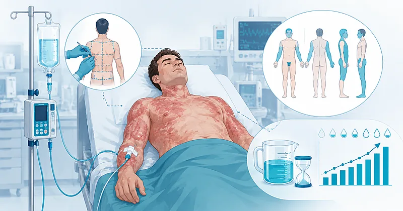
Effective burn fluid resuscitation begins with two measurements taken minutes apart: how much skin is burned, and how deeply. Surface area drives the fluid prescription, depth determines which wounds count, and the patient’s hourly response decides everything that follows. The rules below distil current evidence into actions you can apply from the trauma bay onward.
- 1Estimate surface area with the Lund-Browder chart where available, and exclude simple erythema from the count
- 2Reserve formal burn fluid resuscitation for adults with deep burns of 20% surface area or more
- 3Start at 2 mL of lactated Ringer’s per kg per percent surface area — the current low-volume start point
- 4Start the resuscitation clock at the moment of injury, not at arrival
- 5Titrate hourly to a urine output of 0.5 mL/kg/h — the formula is a starting point, not a target
- 6Watch for fluid creep — cumulative over-resuscitation drives pulmonary and abdominal compartment complications
- 7Consider albumin within the first 24 hours when crystalloid demand outruns the plan, especially in large burns
- 8Identify circumferential full-thickness burns early and escalate for escharotomy before perfusion fails
- 9Refer early to a burn center using accepted criteria — transfer conversations should happen during resuscitation, not after
Estimating Surface Area Before Burn Fluid Resuscitation
Surface-area estimation is the single most consequential number in burn fluid resuscitation, because every downstream fluid calculation scales directly from it. Get it wrong by ten points and you misdose litres of crystalloid. Three bedside tools remain in routine use, each with a different accuracy profile and a different best-fit scenario.
Why estimates drift high
Across reported series, all three traditional methods tend to overestimate, and the error grows with inexperience and with extremes of body habitus. One validation cohort found average overestimates of roughly 8% to 20% surface area depending on the tool used, with the palm method and rule of nines drifting highest. Referring clinicians overestimate more often than burn-center staff, which is one reason a second assessment at the receiving center is standard.
Perform a Lund-Browder assessment for any adult who will need formal resuscitation, since it adjusts for regional body proportions and is the most accurate of the bedside charts.
Strong Rec Moderate Evidence ISBI 2016Use the rule of nines as a rapid prehospital or trauma-bay approximation, then refine the number once the patient is undressed and fully exposed.
Moderate Rec Moderate Evidence ISBI 2016Apply the palmar method only for small or scattered burns, counting the patient’s whole palmar hand surface as roughly 0.8% of total surface area rather than a full 1%.
Conditional Rec Low Evidence ISBI 2016Do not include areas of simple erythema in the surface-area count, since superficial redness does not drive the capillary leak that fluid replaces.
Against High Evidence ISBI 2016Reading Burn Depth at the Bedside
Depth determines which wounds enter the surface-area count and which need surgical attention. Clinical examination of colour, blistering, blanching, and sensation remains the workhorse, though its accuracy for distinguishing superficial from deep partial-thickness wounds is only moderate in the acute window. Most real burns are mixed depth, so describe the dominant pattern rather than forcing a single label.
Depth at a Glance
| Depth | Appearance & Sensation | Counts Toward Surface Area? | Bedside Pitfall |
|---|---|---|---|
| Superficial (epidermal) | Red, dry, painful, blanches briskly; no blisters | No — exclude from the count | Often wrongly added, inflating fluid volume |
| Superficial partial-thickness | Moist, pink, blistered, very painful, blanches | Yes | May deepen over 48h — reassess |
| Deep partial-thickness | Mottled red-white, sluggish or absent blanching, reduced sensation | Yes | Easily mistaken for superficial; often needs grafting |
| Full-thickness | Dry, leathery, white or charred, non-blanching, insensate | Yes | Circumferential limbs threaten perfusion |
Reassess depth at 24 to 48 hours, because superficial partial-thickness wounds can convert to deeper injury and change both the surgical plan and the wound area.
Moderate Rec Low Evidence ISBI 2016Starting Burn Fluid Resuscitation
The starting volume for burn fluid resuscitation has shifted downward over the last decade. Where the classic Parkland figure of 4 mL/kg per percent surface area once anchored practice, the 2024 ABA guidance recommends beginning at half that rate to curb the over-resuscitation that plagued earlier protocols. The number you calculate is an opening bid, not a fixed prescription.
Starting rate at 2 mL/kg per percent: 2 × 80 × 30 = 4,800 mL of lactated Ringer’s estimated for the first 24 hours. Half of that estimate, 2,400 mL, is delivered over the first 8 hours from the time of injury (about 300 mL/h), and the remainder over the following 16 hours. Every figure here is then adjusted hour by hour against urine output.
Initiate burn fluid resuscitation at 2 mL/kg per percent surface area using lactated Ringer’s for adults with deep burns of at least 20% surface area, then titrate upward only as the patient’s response requires.
Strong Rec Moderate Evidence ABA 2024Time the resuscitation from the moment of injury and deliver roughly half the first-day estimate within the first 8 hours, compressing the rate if presentation is delayed.
Strong Rec Moderate Evidence ABLSConsider human albumin within the first 24 hours when crystalloid demand is escalating beyond plan, with a stronger case in larger burns and in rescue scenarios where resuscitation is failing.
Conditional Rec Low Evidence ABA 2024Do not give routine high-dose vitamin C, fresh frozen plasma, or transpulmonary thermodilution-guided targets as standard adjuncts, since current evidence does not support them outside research settings.
Against Moderate Evidence ABA 2024Titration and Resuscitation Endpoints
Urine output is the dominant bedside endpoint. The widely used target for adults is 0.5 mL/kg/h, with most protocols accepting a band of roughly 30 to 50 mL/h in an average-sized adult. The discipline lies in adjusting the infusion in both directions: up when output lags, and just as importantly down when it overshoots.
Titration Logic by Hourly Response
| Hourly Urine Output | Interpretation | Infusion Action | Watch For |
|---|---|---|---|
| Below 0.5 mL/kg/h | Under-resuscitated | Increase rate stepwise; reassess in 1h | Missed surface area; ongoing losses |
| 0.5 to 1 mL/kg/h | On target | Hold rate; continue hourly review | Complacency — keep reassessing |
| Above 1 mL/kg/h | Over-resuscitated | Reduce rate; do not withhold review | Fluid creep; rising intra-abdominal pressure |
Titrate the infusion every hour to a urine output of 0.5 mL/kg/h in adults, treating the calculated formula volume as a reference point rather than a delivery target.
Strong Rec Moderate Evidence ABLSMonitor intra-abdominal and intraocular pressure selectively during large-volume resuscitation, since rising pressures are an early signal of dangerous fluid accumulation.
Strong Rec Moderate Evidence ABA 2024Consider computerised decision-support titration where available, as it may help lower total resuscitation volume by enforcing systematic hourly adjustment.
Conditional Rec Low Evidence ABA 2024Clinical Decision Pathway
A question-based route through the first hours of a major burn. Work the questions in order; each one gates the next.
Referral and Surgical Escalation
Knowing when to involve a burn center, and when a wound needs surgical release, belongs in the resuscitation period rather than after it. The table below pairs each trigger with the action it should prompt.
| Trigger | Why It Matters | Action | Timing |
|---|---|---|---|
| Partial-thickness burn over 10% surface area | Exceeds local management capacity | Refer to burn center | During resuscitation |
| Burns to face, hands, feet, genitalia, or major joints | Functional and cosmetic stakes | Refer to burn center | Early |
| Any full-thickness burn | Likely needs excision and grafting | Refer to burn center | Early |
| Circumferential full-thickness limb or chest burn | Eschar can throttle perfusion or ventilation | Escalate for escharotomy | At first sign of compromise |
| Inhalation injury, chemical, or electrical burn | Atypical fluid and airway course | Refer to burn center | Early |
Refer adults meeting accepted burn center criteria early, and begin the transfer conversation while resuscitation is underway rather than waiting for the patient to stabilise.
Strong Rec Low Evidence ABA Referral CriteriaPerform escharotomy for circumferential full-thickness burns at the earliest sign of vascular or ventilatory compromise, guided by clinical judgement rather than a single objective threshold.
Strong Rec Low Evidence ISBI 2016Monitoring and Follow-Up
| Parameter | When to Check | What to Look For | Common Pitfall |
|---|---|---|---|
| Urine output | Hourly | Steady 0.5 mL/kg/h | Tolerating a high output without cutting the rate |
| Base deficit / lactate | On arrival, then periodically | Trend toward normal as perfusion restores | Chasing a single value instead of the trend |
| Intra-abdominal pressure | When volumes are large or rising | Early rise signalling over-resuscitation | Not measuring until compartment syndrome is overt |
| Distal limb perfusion | Hourly in circumferential burns | Pulses, capillary refill, sensation | Attributing loss of pulse to hypotension alone |
Evidence in Context
Where the major sources converge, where they part company, and how confident the underlying evidence actually is.
From 4 mL/kg to 2 mL/kg: why the start point dropped
The original Parkland figure of 4 mL/kg per percent surface area was a derived estimate, not a ceiling. Decades of practice showed centers routinely exceeding it, and the resulting fluid creep produced pulmonary oedema and abdominal compartment syndrome. The 2024 ABA guidance responded by lowering the recommended start point to 2 mL/kg per percent, framing the calculated volume explicitly as a starting estimate to be titrated down as well as up.
Where the surface-area threshold is genuinely contested
The 20% surface-area threshold for formal resuscitation is widely taught, but it is a pragmatic line rather than a sharp biological one. Some centers resuscitate from 15% to pre-empt acute kidney injury overnight, and recent cohort work has examined outcomes in the 15% to 20% band. The disagreement is about where to draw a continuous risk gradient, not about the underlying physiology.
Albumin and colloid: a measured endorsement
The 2024 ABA review supports considering human albumin to reduce crystalloid volume and improve urine output, with a stronger case in larger burns. Confidence in the evidence is low, and the optimal timing within the first 24 hours remains uncertain. Colloid is best understood as a tool to blunt fluid creep, not a routine first-line agent.
Adjuncts the evidence does not yet support
The same review declined to recommend high-dose vitamin C, fresh frozen plasma, early renal replacement, or vasopressors as routine resuscitation adjuncts, and advised against transpulmonary thermodilution-derived targets. For several of these, the panel could make no recommendation at all because the studies were too small or too few.
References
- 1.Cancio LC, Sheridan RL, Cartotto R, et al. American Burn Association Clinical Practice Guidelines on Burn Shock Resuscitation. J Burn Care Res. 2024;45(3):565–589. doi:10.1093/jbcr/irad125
- 2.ISBI Practice Guidelines Committee. ISBI Practice Guidelines for Burn Care. Burns. 2016;42(5):953–1021. doi:10.1016/j.burns.2016.05.013
- 3.Baxter CR, Shires T. Physiological response to crystalloid resuscitation of severe burns. Ann N Y Acad Sci. 1968;150(3):874–894. doi:10.1111/j.1749-6632.1968.tb14738.x
- 4.Pham TN, Cancio LC, Gibran NS; American Burn Association. American Burn Association practice guidelines: burn shock resuscitation. J Burn Care Res. 2008;29(1):257–266. doi:10.1097/BCR.0b013e31815f3876
How to Read the Evidence Tags
Every recommendation carries two tags — one for recommendation strength and one for evidence quality. These are Medaptly’s own simplified interpretations, not reproductions of any guideline body’s grading system.
Recommendation Strength
| Tag | What It Means |
|---|---|
| Strong Rec | High-quality evidence broadly supports this action. |
| Moderate Rec | The weight of evidence favours this action. |
| Conditional Rec | The benefit is less certain — individualise. |
| Against | Evidence shows no benefit or potential harm. |
Evidence Quality
| Tag | What It Means |
|---|---|
| High Evidence | Multiple well-designed RCTs or high-quality meta-analyses. |
| Moderate Evidence | Single RCT or large observational studies. |
| Low Evidence | Expert consensus or small studies. |