
Idiopathic Intracranial Hypertension: 7 Essential Rules
Clinical Practice Update — Diagnosis, Weight-Based Treatment, Pharmacotherapy, and Surgical Options
This is an original clinical education article informed by current guidelines and evidence. See References below for source documents.
- Clinical Focus
- Evidence-based diagnosis and management of idiopathic intracranial hypertension in adults
- Target Audience
- Primary care, neurologists, ophthalmologists, neurosurgeons, emergency physicians
- Setting
- Primary care, neurology outpatient, emergency department, neuro-ophthalmology clinic
- Source Evidence
- •Friedman et al. Revised Diagnostic Criteria for Pseudotumor Cerebri Syndrome (Neurology, 2013)
- •Mollan et al. IIH Consensus Guidelines on Management (JNNP, 2018)
- •Wall et al. NORDIC IIHTT — Acetazolamide Trial (JAMA, 2014)
- •Hoffmann et al. European Headache Federation Guideline on IIH
- •Mollan et al. IIH:WT Weight-Loss Study (JAMA Neurology, 2021)
Key Clinical Takeaways
Effective management of idiopathic intracranial hypertension depends on three parallel priorities: protect vision, relieve headache, and drive sustained weight loss. The modified Dandy criteria (Friedman 2013) define who has the condition; the NORDIC trial defined what to do medically; the IIH:WT study cemented that weight loss is the single most powerful disease-modifying intervention. The points below distil that evidence into actionable rules for everyday practice.
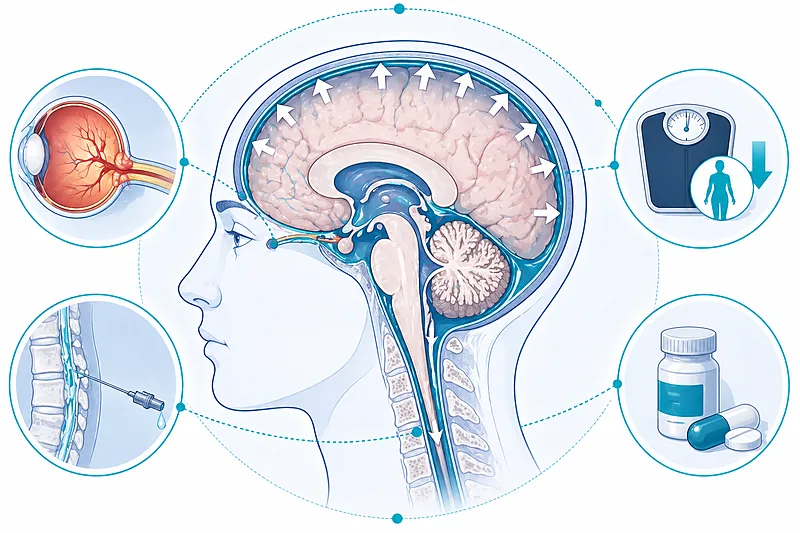
- 1Apply the modified Dandy criteria (Friedman 2013) to confirm idiopathic intracranial hypertension — papilledema, normal neuro-exam aside from cranial nerve VI palsy, neuroimaging showing no alternative cause, and elevated LP opening pressure with normal CSF composition → Diagnosis
- 2Always order MRI with MR venography to exclude cerebral venous sinus thrombosis before accepting a diagnosis of idiopathic intracranial hypertension → Diagnosis
- 3Measure LP opening pressure with the patient relaxed in the lateral decubitus position — > 25 cm H₂O (adults) or > 28 cm H₂O (children) supports the diagnosis → Diagnosis
- 4Refer every patient for urgent neuro-ophthalmology review at diagnosis for formal perimetry and optical coherence tomography baseline → Initial Management
- 5Make sustained weight loss the cornerstone of management — a 5–10% reduction in body weight can induce remission in idiopathic intracranial hypertension → Initial Management
- 6Start acetazolamide as first-line medical therapy, titrated to tolerance — NORDIC trial data support doses up to 4 g/day with supervised escalation → Medical Treatment
- 7Escalate to surgical intervention — CSF shunting, venous sinus stenting, or optic nerve sheath fenestration — when vision is threatened despite optimal medical therapy → Surgical
- 8Treat headache as a separate clinical problem — resolution of raised pressure does not guarantee resolution of headache, and migraine-type headache is common → Monitoring
- 9Monitor vision throughout — serial perimetry and OCT are the most sensitive tools to detect early deterioration before irreversible optic nerve damage occurs → Monitoring
Diagnosing Idiopathic Intracranial Hypertension
Idiopathic intracranial hypertension is a diagnosis of exclusion built on three pillars: a consistent clinical picture, confirmatory lumbar puncture, and neuroimaging that rules out alternative causes of raised intracranial pressure. The Friedman 2013 revision of the Dandy criteria has become the practical standard; it balances sensitivity with the need to reliably exclude secondary causes of raised pressure such as venous sinus thrombosis, mass lesion, or drug-induced intracranial hypertension.
Typical Clinical Features
The classical presentation is daily headache (often pulsatile, worse on waking, aggravated by Valsalva manoeuvres and lying flat), pulsatile tinnitus, and transient visual obscurations — brief blackouts of vision lasting seconds, provoked by postural change or coughing. Diplopia from cranial nerve VI palsy occurs in a minority. The archetypal patient is a young woman with obesity, though the condition can occur in any demographic and must not be dismissed because the patient doesn’t “look typical.”
Perform dilated fundoscopy in every patient suspected of idiopathic intracranial hypertension. Papilledema is the cardinal finding; document grade using the Frisén scale, bilateral asymmetry, haemorrhages, or cotton wool spots.
Strong Rec High Evidence Friedman 2013 Mollan 2018Order MRI of the brain with MR venography as the first-line imaging study. MRV is essential to exclude cerebral venous sinus thrombosis, which presents almost identically and is the single most important mimic to exclude before labelling a patient with idiopathic intracranial hypertension.
Strong Rec High Evidence Mollan 2018 EHFPerform lumbar puncture with the patient in lateral decubitus position, legs relaxed and extended. Opening pressure > 25 cm H₂O in adults (or > 28 cm H₂O in children with sedation) supports the diagnosis. Send CSF for cell count, protein, glucose, and cytology to confirm normal composition.
Strong Rec High Evidence Friedman 2013Review the medication list for secondary causes before confirming idiopathic intracranial hypertension. Tetracyclines (doxycycline, minocycline), vitamin A and retinoids, lithium, prolonged systemic steroids, and growth hormone have all been implicated. Withdrawal of the offending agent may be curative.
Strong Rec Moderate Evidence Friedman 2013 Mollan 2018Supportive MRI Findings
| Sign | Where to Look | Clinical Value | Practical Tip |
|---|---|---|---|
| Empty sella | Sagittal T1 pituitary fossa | Highly specific; common in chronic disease | Not sufficient alone; most useful alongside other signs |
| Posterior globe flattening | Axial T2 through the orbits | Specific to elevated pressure | Request dedicated orbital cuts if clinical suspicion is high |
| Distended optic nerve sheath | Axial T2, orbit | Supports diagnosis; can be measured | Compare with the fellow eye for subtle asymmetry |
| Transverse sinus stenosis | MRV, 2D TOF and contrast sequences | Relevant for venous sinus stenting candidacy | Distinguish from CVST — stenosis, not thrombus |
| Protruding optic nerve head | Axial T2 orbit | Corresponds to papilledema on fundoscopy | Correlates with visual risk |
Initial Management of Idiopathic Intracranial Hypertension
Once the diagnosis is secure, initial management of idiopathic intracranial hypertension sits on three legs: baseline vision assessment, a weight-loss plan that is both realistic and sustained, and medical therapy matched to disease severity. Triage is urgent when the patient has fulminant disease — rapidly progressive visual loss over days — which warrants inpatient admission and usually immediate surgical consultation.
Refer for formal neuro-ophthalmology assessment at the time of diagnosis. Baseline automated perimetry (Humphrey 24-2 or 30-2), OCT retinal nerve fibre layer thickness, and colour vision testing are essential to detect subsequent deterioration before it becomes symptomatic.
Strong Rec High Evidence Mollan 2018Counsel every patient with idiopathic intracranial hypertension on the central role of weight loss. A sustained 5–10% reduction in body weight reliably reduces intracranial pressure and papilledema, and can induce clinical remission. Frame weight loss as the primary disease-modifying treatment — not as a lifestyle aside.
Strong Rec High Evidence IIH:WT 2021 Mollan 2018Consider bariatric surgery in patients with idiopathic intracranial hypertension and BMI ≥ 35 kg/m² where conservative weight-loss efforts have failed. The IIH:WT trial showed bariatric surgery was superior to community weight-management programmes for both intracranial pressure and papilledema.
Moderate Rec High Evidence IIH:WT 2021Recognise and triage fulminant idiopathic intracranial hypertension separately. Rapidly progressive vision loss (days, not weeks), severe papilledema, or profound headache warrant emergency admission, urgent repeat LP or shunting, and neuro-ophthalmology involvement within hours.
Strong Rec Moderate Evidence Mollan 2018Medical Treatment Options
Pharmacotherapy for idiopathic intracranial hypertension supports the weight-loss plan rather than replacing it. Acetazolamide is the first-line agent, supported by the NORDIC IIHTT trial. Topiramate is a reasonable alternative for patients with coexisting migraine or those who do not tolerate acetazolamide. Furosemide is a third option, often used as an add-on. Optimal dosing matters: under-dosing is a common cause of apparent treatment failure.
Start acetazolamide 250–500 mg twice daily as first-line pharmacotherapy for idiopathic intracranial hypertension. Titrate by 250–500 mg weekly as tolerated. NORDIC-trial patients tolerated doses up to 4 g/day; target the highest dose the patient can tolerate.
Strong Rec High Evidence NORDIC 2014 Mollan 2018Counsel patients on the side-effect profile of acetazolamide: paraesthesiae (common, often limiting), altered taste (especially of carbonated drinks), fatigue, and renal stones. Monitor serum bicarbonate, potassium, and creatinine at baseline, 2–4 weeks, then every 3–6 months.
Strong Rec Moderate Evidence Mollan 2018Consider topiramate (titrated to 100–200 mg/day in divided doses) when acetazolamide is not tolerated, or preferentially when migraine-type headache is a prominent feature. The weight-loss side effect is welcome in this patient group.
Moderate Rec Moderate Evidence Mollan 2018 EHFConsider furosemide (20–40 mg twice daily) as an add-on when acetazolamide alone is insufficient, or as monotherapy when carbonic anhydrase inhibitors are contraindicated. Evidence is weaker than for acetazolamide but clinically established.
Conditional Rec Low Evidence Mollan 2018Avoid systemic corticosteroids except as a short bridge to definitive treatment in fulminant idiopathic intracranial hypertension. Rebound intracranial hypertension on withdrawal, weight gain, and metabolic side effects make them unsuitable for chronic use.
Against Moderate Evidence Mollan 2018Treat associated headache as a distinct problem. A substantial proportion of patients continue to have migraine-pattern headache even after intracranial pressure normalises; standard migraine preventives and acute therapies should be applied accordingly.
Strong Rec Moderate Evidence Mollan 2018 EHFPharmacological Options: A Drug-by-Drug Guide
| Drug | Starting Dose | Target Range | Key Monitoring | Practical Tips |
|---|---|---|---|---|
| Acetazolamide | 250–500 mg BD | 1–4 g/day divided | Bicarbonate, potassium, creatinine | Paraesthesiae settle in weeks; taste changes persist; pregnancy category risk discussion needed |
| Topiramate | 25 mg nightly | 100–200 mg/day | Cognitive effects, renal stones | Helpful when migraine coexists; slow titration to minimise cognitive side effects |
| Furosemide | 20 mg BD | 40–80 mg/day | Electrolytes, blood pressure | Useful as add-on; weaker evidence; risk of hypokalaemia |
| Corticosteroids | N/A (short bridge only) | Not for chronic use | Glucose, weight, blood pressure | Reserved for fulminant disease as bridge to surgery |
Surgical and Interventional Options in Idiopathic Intracranial Hypertension
Surgical treatment of idiopathic intracranial hypertension is indicated when vision is threatened despite optimal medical therapy, when fulminant disease is present at diagnosis, or when intractable headache fails to respond to weight loss and pharmacotherapy. Three main approaches are available: CSF diversion (shunting), venous sinus stenting, and optic nerve sheath fenestration. Each has distinct indications, advantages, and failure patterns.
Refer urgently for CSF shunting (ventriculoperitoneal or lumboperitoneal) when there is progressive visual loss despite maximal tolerated medical therapy or fulminant disease at presentation. Ventriculoperitoneal shunts have lower revision rates than lumboperitoneal in most modern series.
Strong Rec Moderate Evidence Mollan 2018Consider venous sinus stenting in patients with documented significant transverse sinus stenosis and a trans-stenotic pressure gradient ≥ 8–10 mmHg. Stenting can normalise pressure, reduce papilledema, and improve headache, with outcomes comparable to shunting in selected series.
Moderate Rec Moderate Evidence Mollan 2018Consider optic nerve sheath fenestration when the primary threat is to vision and headache is not a dominant feature. Fenestration addresses papilledema and optic nerve oedema but does not reliably relieve headache or normalise intracranial pressure.
Moderate Rec Low Evidence Mollan 2018Counsel patients that surgical intervention does not remove the need for weight loss. Recurrence is common in patients who regain weight or fail to achieve sustained weight reduction after the procedure.
Strong Rec Moderate Evidence Mollan 2018Comparing Surgical Options
| Procedure | Best Candidate | Primary Benefit | Main Trade-Offs |
|---|---|---|---|
| Ventriculoperitoneal shunt | Refractory disease, fulminant vision loss | Rapid pressure reduction; improves vision and headache | Shunt failure/revision common; infection risk; over-drainage headaches |
| Lumboperitoneal shunt | Historically first-line; now less favoured | Avoids ventricular puncture | Higher revision rate; tonsillar herniation risk if over-drainage |
| Venous sinus stenting | Documented sinus stenosis with pressure gradient | Normalises pressure without implanted shunt; reduces headache | Needs specialist centre; lifelong antiplatelets; adjacent-segment stenosis |
| Optic nerve sheath fenestration | Vision-threatening papilledema, minimal headache | Directly protects the optic nerve | Does not address headache; may need bilateral procedures |
Clinical Decision Pathway
A practical, question-based walk-through of suspected idiopathic intracranial hypertension from first presentation to definitive management. Work through the questions in order.
Monitoring and Follow-Up
Ongoing care in idiopathic intracranial hypertension is shared between neurology, neuro-ophthalmology, and primary care. Four domains should be tracked at every review: vision, papilledema, headache, and weight. Disease may relapse years after apparent remission, particularly with weight regain, so patients should be counselled to return at any point with recurring symptoms.
| Parameter | When to Check | What to Look For | Common Pitfalls |
|---|---|---|---|
| Automated perimetry | Baseline; every 1–3 months initially; every 6 months once stable | Enlarging blind spot, arcuate defects, nasal step, generalised depression | Relying on confrontation fields — too insensitive for early loss |
| Optical coherence tomography | Baseline; at each neuro-ophthalmology visit | Peripapillary RNFL thickness trend; GCL-IPL thinning signals axon loss | Mistaking resolving oedema for atrophy — correlate with exam |
| Papilledema grading | Every visit with dilated exam | Frisén grade, bilateral asymmetry, haemorrhages | Missing subtle resolution of swelling with emerging pallor (optic atrophy) |
| Headache diary | Ongoing | Frequency, severity, medication overuse | Attributing all headache to raised pressure once pressure has normalised |
| Weight and BMI | Every visit | Sustained weight change (positive or negative) | Discussing weight without concrete support plan is rarely effective |
| Bicarbonate / electrolytes | Baseline, 2–4 weeks, then every 3–6 months on acetazolamide | Metabolic acidosis, hypokalaemia | Stopping the drug for mild chemistry changes alone |
Evidence in Context
What the current evidence supports, where the major guidelines align, and where clinically relevant gaps remain in the management of idiopathic intracranial hypertension.
Where UK Consensus and European Guidelines Align
Both the Mollan 2018 UK consensus and the European Headache Federation guideline converge on the central principles: the Friedman 2013 criteria define the condition, weight loss is the primary disease-modifying intervention, acetazolamide is first-line pharmacotherapy, and urgent surgical intervention is indicated for vision-threatening disease. Both also emphasise the importance of treating headache as a separate problem and recognising fulminant disease as an emergency.
NORDIC IIHTT: What Acetazolamide Actually Does
The NORDIC Idiopathic Intracranial Hypertension Treatment Trial randomised patients with mild vision loss to acetazolamide (up to 4 g/day) plus a dietary weight-loss programme versus placebo plus diet. The acetazolamide arm showed greater improvement in perimetric mean deviation, reduced papilledema, and lower intracranial pressure. The effect was clinically meaningful, supporting dose escalation as tolerated rather than under-dosing.
IIH:WT: Bariatric Surgery vs Community Weight Management
The IIH:WT randomised trial compared bariatric surgery with a structured community weight-loss programme in women with idiopathic intracranial hypertension and BMI above 35 kg/m². Bariatric surgery produced substantially greater weight loss, bigger reductions in intracranial pressure, and better papilledema outcomes at 12 and 24 months. The trial reframed weight-loss surgery from last-resort option to evidence-based first-line intervention in this specific subgroup.
Stenting vs Shunting: The Unresolved Comparison
Observational series suggest venous sinus stenting is at least as effective as CSF diversion in appropriately selected patients with transverse sinus stenosis and significant pressure gradient. Head-to-head randomised comparisons are not yet available. Until they are, choice remains driven by local expertise, anatomy, and patient preference. A multidisciplinary discussion is appropriate for every surgical candidate.
What We Still Don’t Know
The precise pathophysiology of idiopathic intracranial hypertension remains incompletely characterised, though androgen metabolism, venous outflow anatomy, and obesity-related mechanisms are all implicated. Novel therapeutic targets such as GLP-1 receptor agonists for concurrent weight loss are under active investigation. Predictors of treatment failure and best practice for long-term monitoring after remission remain open questions.
References
- 1.Friedman DI, Liu GT, Digre KB. Revised diagnostic criteria for the pseudotumor cerebri syndrome in adults and children. Neurology. 2013;81(13):1159–1165. doi:10.1212/WNL.0b013e3182a55f17
- 2.Mollan SP, Davies B, Silver NC, et al. Idiopathic intracranial hypertension: consensus guidelines on management. J Neurol Neurosurg Psychiatry. 2018;89(10):1088–1100. doi:10.1136/jnnp-2017-317440
- 3.Wall M, McDermott MP, Kieburtz KD, et al. Effect of acetazolamide on visual function in patients with idiopathic intracranial hypertension and mild visual loss: the idiopathic intracranial hypertension treatment trial. JAMA. 2014;311(16):1641–1651. doi:10.1001/jama.2014.3312
- 4.Mollan SP, Mitchell JL, Ottridge RS, et al. Effectiveness of Bariatric Surgery vs Community Weight Management Intervention for the Treatment of Idiopathic Intracranial Hypertension: A Randomized Clinical Trial. JAMA Neurol. 2021;78(6):678–686. doi:10.1001/jamaneurol.2021.0659
- 5.Hoffmann J, Mollan SP, Paemeleire K, et al. European Headache Federation guideline on idiopathic intracranial hypertension. J Headache Pain. 2018;19(1):93. doi:10.1186/s10194-018-0919-2
- 6.Markey KA, Mollan SP, Jensen RH, Sinclair AJ. Understanding idiopathic intracranial hypertension: mechanisms, management, and future directions. Lancet Neurol. 2016;15(1):78–91. doi:10.1016/S1474-4422(15)00298-7
How to Read the Evidence Tags
Every recommendation in this article carries two tags — one for recommendation strength and one for evidence quality — using Medaptly’s own simplified interpretations.
Recommendation Strength
| Tag | What It Means |
|---|---|
| Strong Rec | High-quality evidence broadly supports this action. |
| Moderate Rec | The weight of evidence favours this action. |
| Conditional Rec | The benefit is less certain — individualise based on patient factors. |
| Against | Evidence shows no benefit or potential harm. |
Evidence Quality
| Tag | What It Means |
|---|---|
| High Evidence | Multiple well-designed RCTs or high-quality meta-analyses. |
| Moderate Evidence | Single RCT or large observational studies. |
| Low Evidence | Expert consensus or small studies. |