
Inclusion Body Myositis: A Diagnostic and Supportive Care Framework
Clinical Practice Update — Pattern of Weakness, Biopsy Findings, and Physical Therapy Focus
This is an original clinical education article informed by current guidelines and evidence. See References below for source documents.
- Clinical Focus
- Recognition, diagnosis, and supportive care of inclusion body myositis in adults
- Target Audience
- Neurologists, rheumatologists, general internists, primary care physicians, physical therapists
- Setting
- Outpatient neurology and primary care, neuromuscular referral clinics
- Source Evidence
- •ENMC International Workshop Diagnostic Criteria for IBM (2011, updated 2024)
- •Lloyd TE et al. Evaluation and construction of diagnostic criteria for IBM. Brain (2014)
- •Naddaf E, Barohn RJ, Dimachkie MM. IBM: Update on Pathogenesis and Treatment. Neurotherapeutics (2018)
- •Greenberg SA. Inclusion body myositis: clinical features and pathogenesis. Nat Rev Rheumatol (2019)
Key Clinical Takeaways
Inclusion body myositis is the most common acquired myopathy in adults over 50, yet it is routinely misdiagnosed as polymyositis for years. Three features anchor a confident diagnosis: a characteristic weakness pattern (finger flexors and quadriceps), supportive muscle biopsy findings, and a disease course that fails to respond to immunosuppression. Because no disease-modifying therapy works in inclusion body myositis, an effective supportive care plan — built around physical therapy, dysphagia management, and falls prevention — is where clinical value is generated.
- 1Suspect inclusion body myositis in any adult over 50 with slowly progressive, often asymmetric weakness involving the finger flexors and quadriceps
- 2Test deep finger flexor strength specifically — weak grip with relatively spared shoulder abduction is highly suggestive
- 3Reconsider inclusion body myositis whenever a presumed polymyositis case fails to improve on steroids
- 4Creatine kinase is usually mildly elevated (under 15× ULN) and may be normal — do not exclude IBM on a low CK
- 5Muscle biopsy classically shows rimmed vacuoles, endomysial inflammation invading non-necrotic fibers, and COX-negative fibers
- 6cN-1A (anti-NT5C1A) antibodies are specific for inclusion body myositis but only positive in roughly one-third to half of patients
- 7Muscle MRI shows a remarkably selective pattern — distal vastus medialis and forearm flexor compartment fatty replacement
- 8Multiple trials have failed to show benefit from prednisone, methotrexate, IVIG, or biologics — chronic immunosuppression is not warranted
- 9Refer every patient to a neuromuscular-trained physical therapist for individualised aerobic and resistance training
- 10Screen for dysphagia at every visit — aspiration pneumonia is the leading cause of disease-related mortality
Recognizing the Pattern: Inclusion Body Myositis at the Bedside
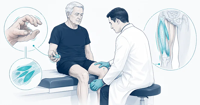
The clinical pattern of inclusion body myositis is its most reliable diagnostic clue, and most patients are misdiagnosed because the pattern is not specifically sought. The disease produces a distinctive asymmetric weakness that preferentially affects two muscle groups: the deep finger flexors of the forearm and the quadriceps. Recognising this pattern requires deliberate examination — weakness of these muscles is easy to miss if the examiner only screens proximal shoulder and hip muscles.
Patients typically present in their sixth or seventh decade, with men affected roughly two to three times more often than women. The earliest functional complaints are difficulty rising from low chairs, frequent tripping or knee buckling, and a progressive inability to grip — opening jars, turning keys, or buttoning shirts becomes effortful. Symptoms creep on so insidiously that patients often date onset to a specific fall or injury when in fact the underlying weakness has been progressing for years.
Suspect inclusion body myositis in any adult over 50 presenting with slowly progressive, asymmetric weakness of the finger flexors and quadriceps. The combination is highly specific.
Strong Rec High Evidence ENMC 2024Test deep finger flexion specifically. Ask the patient to flex the distal interphalangeal joints against resistance — weakness here, with relatively spared shoulder abduction, is the bedside hallmark of inclusion body myositis.
Strong Rec Moderate Evidence Greenberg 2019Inspect the forearms for selective atrophy of the volar compartment, and the thighs for vastus medialis wasting. Visible atrophy in these regions in an older adult with weakness is strongly suggestive.
Moderate Rec Moderate Evidence Greenberg 2019Evaluate for facial weakness and mild dysphagia at presentation. Both are common in inclusion body myositis and often unreported by patients.
Moderate Rec Moderate Evidence Cox 2011Reconsider the diagnosis of polymyositis whenever a patient fails to improve on adequate corticosteroid therapy. Many cases labelled steroid-refractory polymyositis are actually inclusion body myositis.
Strong Rec High Evidence ENMC 2024Clinical Pattern of Weakness: What to Examine
| Body Region | Affected Muscles | Functional Impact | Bedside Test |
|---|---|---|---|
| Forearm (volar) | Flexor digitorum profundus, flexor pollicis longus | Weak grip, dropping objects, difficulty with keys and buttons | Resisted distal IP joint flexion of all fingers simultaneously |
| Thigh (anterior) | Quadriceps — especially distal vastus medialis | Knee buckling, falls, difficulty rising from chairs and stairs | Resisted knee extension; observe sit-to-stand |
| Lower leg | Tibialis anterior, medial gastrocnemius | Foot drop, tripping, calf wasting | Heel walking; check for asymmetric ankle dorsiflexion |
| Face | Orbicularis oculi, lower face | Mild facial weakness, often unrecognised by patient | Resisted eye closure and cheek puff |
| Pharynx | Pharyngeal constrictors, cricopharyngeus | Dysphagia, coughing with thin liquids, weight loss | Targeted swallowing questions (EAT-10 screen) |
Muscle Biopsy Findings in Inclusion Body Myositis
Muscle biopsy remains a central step in confirming inclusion body myositis, but the findings must be interpreted by a pathologist familiar with neuromuscular pathology and correlated with the clinical pattern. No single feature is both sensitive and specific — the diagnosis rests on a constellation of findings, and absence of one classic feature does not exclude the disease.
Choose a muscle biopsy site that is moderately affected — not end-stage atrophic and not unaffected. Open biopsy of the vastus lateralis or biceps brachii is conventional; needle biopsy is acceptable in experienced hands but yields less tissue. Tissue should be processed for routine histology, enzyme histochemistry, immunohistochemistry, and electron microscopy.
Perform open muscle biopsy from a moderately affected muscle — typically vastus lateralis or biceps brachii. Avoid severely atrophic muscle where fibrofatty replacement may obscure findings.
Strong Rec High Evidence ENMC 2024Examine for endomysial inflammation with CD8+ T-cell invasion of non-necrotic, MHC-I-expressing muscle fibers — the inflammatory hallmark of inclusion body myositis.
Strong Rec High Evidence ENMC 2024Look for rimmed vacuoles on modified Gomori trichrome — basophilic granular material lining vacuolar membranes. Their presence strongly supports the diagnosis but they may be absent in early disease.
Strong Rec High Evidence ENMC 2024Quantify cytochrome c oxidase (COX) negative fibers — an increased proportion above age-matched controls supports inclusion body myositis and reflects mitochondrial dysfunction characteristic of the disease.
Strong Rec Moderate Evidence Brady 2014Request p62 and TDP-43 immunostaining when available. Cytoplasmic aggregates of these proteins are sensitive markers and may be positive when rimmed vacuoles are not yet visible.
Moderate Rec Moderate Evidence Hiniker 2013Do not exclude inclusion body myositis when rimmed vacuoles are absent. A clinical pattern that is otherwise characteristic, combined with inflammation and COX-negative fibers, is sufficient under current criteria for clinically defined disease.
Moderate Rec Moderate Evidence Lloyd 2014Hierarchy of Biopsy Findings: How to Read the Report
| Finding | Detection Method | Diagnostic Weight | When Absent |
|---|---|---|---|
| Endomysial CD8+ T-cells invading non-necrotic fibers | CD8 immunostain + MHC-I overexpression | High — required for pathological diagnosis | Reconsider non-inflammatory myopathy |
| Rimmed vacuoles | Modified Gomori trichrome | High when present; absence does not exclude | Diagnosis can still stand on clinical grounds |
| COX-negative fibers | COX/SDH double stain | Moderate; supportive when increased above age-norms | Less weighty in early disease |
| p62 / TDP-43 aggregates | Immunohistochemistry | Moderate; supports diagnosis when rimmed vacuoles absent | Often not done routinely — request specifically |
| 15–18 nm tubulofilamentous inclusions | Electron microscopy | Highly specific when found | Sampling-limited; reserved for difficult cases |
Antibodies, Imaging, and Differential Diagnosis
When the clinical pattern and biopsy converge, the diagnosis is usually secure. When they do not, three additional tools refine the diagnosis: serum antibody testing, muscle MRI, and a deliberate review of the differential.
Anti-cN-1A (anti-NT5C1A) antibodies are the only widely available serological marker for inclusion body myositis. Sensitivity is roughly 33 to 50 percent and specificity against other inflammatory myopathies is around 90 to 95 percent. A positive result substantially raises diagnostic confidence; a negative result does not exclude the disease. Muscle MRI shows a strikingly selective pattern of fatty replacement — the distal vastus medialis is often affected disproportionately, and forearm flexor compartment involvement is rarely seen in other myopathies.
Send cN-1A (anti-NT5C1A) antibody testing in suspected inclusion body myositis. A positive result supports the diagnosis; a negative result does not exclude it.
Moderate Rec Moderate Evidence Lloyd 2016Order muscle MRI of the thighs and forearms when the diagnosis is uncertain. Selective fatty replacement of the distal vastus medialis and the forearm flexor compartment is highly characteristic.
Moderate Rec Moderate Evidence Cox 2011Perform EMG to confirm a myopathic process with superimposed neurogenic features — long-duration units, fibrillations, and positive sharp waves are common in inclusion body myositis.
Strong Rec Moderate Evidence ENMC 2024Check creatine kinase but do not rely on it. CK in inclusion body myositis is typically only mildly elevated (often under 1500 U/L) and may be normal — a low CK does not refute the diagnosis.
Moderate Rec Moderate Evidence Naddaf 2018Differential Diagnosis: Conditions That Mimic Inclusion Body Myositis
| Condition | Distinguishing Feature | How to Test | Why It Matters |
|---|---|---|---|
| Polymyositis | Symmetric proximal weakness; higher CK; responds to steroids | Examine finger flexors; trial of steroids | Most common misdiagnosis; immunosuppression exposure |
| Motor neuron disease (ALS) | Upper motor neuron signs; fasciculations; bulbar onset | Neurological exam; EMG showing diffuse denervation | Faster progression; very different prognosis |
| Necrotizing autoimmune myopathy | High CK; proximal weakness; anti-HMGCR or anti-SRP antibodies | Myositis antibody panel | Treatable with immunosuppression |
| Late-onset muscular dystrophy | Family history; no inflammation on biopsy | Genetic panel; biopsy review | Genetic counselling implications |
| Cervical myelopathy | Sensory level; hyperreflexia; bladder involvement | MRI cervical spine | Surgically correctable |
Why Inclusion Body Myositis Resists Immunosuppression
Despite the prominent endomysial inflammation on biopsy, inclusion body myositis does not behave like an immune-mediated disease in trials. Prednisone, methotrexate, azathioprine, mycophenolate mofetil, intravenous immunoglobulin, rituximab, alemtuzumab, and bimagrumab have all been studied. None has produced sustained functional improvement.
The contemporary view is that inclusion body myositis combines an inflammatory component with a degenerative process — protein aggregation, autophagy dysfunction, and mitochondrial damage — that progresses independently of inflammation. Suppressing the immune system addresses only one pathway and does not slow the disease. Sirolimus has shown signals of efficacy in a small Phase 2b trial through enhanced autophagy rather than immunosuppression and is the subject of ongoing study; the trial pipeline remains active but no agent has yet earned routine use.
Do not initiate chronic corticosteroids, methotrexate, IVIG, or biologics for typical inclusion body myositis. Multiple controlled trials show no meaningful functional benefit and substantial cumulative harm.
Against High Evidence Multiple RCTsConsider a short, time-limited trial of immunotherapy (typically 3 months of prednisone with objective strength measurement) only when the diagnosis is genuinely ambiguous after biopsy — not in clinically and pathologically confirmed disease.
Conditional Rec Low Evidence Expert ConsensusRefer interested patients to active clinical trials. Novel agents targeting autophagy, protein aggregation, and muscle anabolic pathways remain under investigation.
Conditional Rec Low Evidence Expert ConsensusSupportive Care: The Physical Therapy Focus
Because no drug modifies the natural history of inclusion body myositis, the supportive care plan is where clinical value is produced. The most consistently studied and effective intervention is structured exercise. Patients with inclusion body myositis who maintain physical activity preserve function longer than those who do not, and the long-held fear that exercise might worsen inflammation has not been borne out in any controlled study.
The right referral matters. A neuromuscular physical therapy program will look different from a generic outpatient program: it emphasises aerobic capacity, eccentric and concentric resistance training of muscles that are not severely weak, range-of-motion preservation in affected joints, and an explicit functional goal (e.g., independent transfers, safe ambulation). Eccentric training in particular — controlled lowering against gravity — produces strength gains in inclusion body myositis when concentric training plateaus.
Refer every patient with inclusion body myositis to a physical therapist with neuromuscular experience for an individualised exercise prescription. The exercise plan, not a drug, is the single most important intervention.
Strong Rec Moderate Evidence Jensen 2019Prescribe aerobic exercise — stationary cycling, recumbent bicycle, or pool walking — three to five times per week at moderate intensity. Cardiovascular fitness improves measurably with no evidence of muscle harm.
Strong Rec Moderate Evidence Spector 1997Add resistance training targeting muscles graded 4 or stronger on MRC scale. Submaximal loading two to three times per week produces measurable strength gains without precipitating muscle damage.
Moderate Rec Moderate Evidence Arnardottir 2003Counsel patients clearly that disuse atrophy is preventable and that bed rest worsens function. Many patients reduce activity after diagnosis out of fear — this accelerates decline.
Strong Rec Moderate Evidence Jensen 2019Assess falls risk at every visit. Fit an ankle-foot orthosis early for foot drop and offer knee bracing or a wheeled walker before a serious fall, rather than after.
Strong Rec Moderate Evidence Jensen 2019Refer to occupational therapy for hand function preservation. Built-up utensils, key turners, button hooks, and adapted clothing fasteners preserve independence as finger flexor weakness progresses.
Moderate Rec Low Evidence Expert ConsensusPhysical Therapy Prescription: What to Recommend
| Exercise Type | Recommended Approach | Frequency | What to Avoid |
|---|---|---|---|
| Aerobic | Stationary cycling, recumbent bike, pool walking, treadmill at moderate pace | 3–5 sessions per week, 20–40 min | Long sedentary periods; deconditioning after illness |
| Resistance (concentric/eccentric) | Submaximal loading of MRC 4+ muscles, including controlled eccentric movements | 2–3 sessions per week, 8–12 reps | Maximal effort to fatigue on severely weak muscles |
| Balance and gait training | Standing balance progression; tandem walking; dual-task practice | Daily home practice + weekly PT | Unsupervised high-fall-risk activities |
| Range of motion | Daily passive and active stretching, especially heel cords and finger flexors | Daily | Allowing contractures to develop unchallenged |
| Functional task practice | Sit-to-stand reps; stair practice with rail; grip and key turning | Daily, integrated into routines | Avoiding tasks because they have become harder |
Managing Dysphagia and Falls
Dysphagia affects roughly 40 to 65 percent of patients with inclusion body myositis and is the leading source of disease-related mortality through aspiration pneumonia. It is also one of the most under-recognised features — patients adapt their diets quietly and may not volunteer symptoms unless asked specifically.
Screen with a validated tool such as the EAT-10 questionnaire at every clinic visit. Any positive screen or report of coughing with thin liquids, food sticking in the throat, or unintentional weight loss warrants formal evaluation with a video fluoroscopic swallow study. The most common physiologic problem is cricopharyngeal dysfunction — failure of the upper oesophageal sphincter to relax. Selected patients with cricopharyngeal obstruction benefit from balloon dilation or cricopharyngeal myotomy.
Screen for dysphagia at every visit using a validated tool. Ask specifically about coughing with thin liquids, food sticking, and unintentional weight loss.
Strong Rec Moderate Evidence Naddaf 2018Refer for video fluoroscopic swallow study at symptom onset, with re-evaluation as swallowing function changes. Diet modification and posture training reduce aspiration risk.
Strong Rec Moderate Evidence Cox 2009Consider cricopharyngeal balloon dilation or myotomy for selected patients with cricopharyngeal dysfunction documented on swallow study. Benefit is often substantial but temporary — repeat procedures may be required.
Conditional Rec Moderate Evidence Oh 2007Vaccinate against influenza and pneumococcal disease per age-appropriate schedules. Aspiration pneumonia is the most common cause of disease-related mortality.
Strong Rec High Evidence CDC 2024Discuss prognosis honestly. Inclusion body myositis is slowly progressive; most patients require ambulatory assistance within 10 to 15 years of symptom onset. Life expectancy is generally only modestly reduced, primarily through aspiration risk.
Strong Rec Moderate Evidence Cox 2011Address mood and social isolation. Depression and reduced social engagement are common as functional independence declines — ask about both and refer when indicated.
Moderate Rec Low Evidence Expert ConsensusClinical Decision Pathway
A practical, question-based approach to working up suspected inclusion body myositis from first presentation to long-term care plan.
Monitoring and Follow-Up
Long-term care emphasises objective tracking of function rather than chasing laboratory markers. The goals are to detect functional decline early, intervene before complications, and maintain independence as long as possible.
| Parameter | When to Check | What to Look For | Common Pitfalls |
|---|---|---|---|
| Functional measures (mIBMFRS, 6-minute walk) | Every 6–12 months | Decline in transfers, gait, ADLs, swallowing | Relying on patient self-report alone |
| Manual muscle testing (MMT) | Every visit | Asymmetric decline; new muscle group involvement | Skipping finger flexors and quadriceps in busy clinics |
| Dysphagia screen | Every visit | New coughing, weight loss, pneumonia history | Patients minimise symptoms; ask family directly |
| Falls history | Every visit | Number, mechanism, injuries since last visit | Underreporting from embarrassment |
| CK | At diagnosis and as clinically indicated | No need to recheck routinely — does not track disease activity | Over-ordering creates false reassurance or alarm |
| Weight, BMI, nutrition | Every visit | Unintentional weight loss may herald advancing dysphagia | Sarcopenia attributed to age rather than disease |
Evidence in Context
What the evidence base supports, where diagnostic criteria differ, and where the literature on treatment stands.
How ENMC 2024 Differs from Earlier Criteria
The 2024 update of the ENMC criteria places greater weight on the clinical pattern of finger flexor and quadriceps weakness, recognising that older criteria over-relied on rimmed vacuoles. Under the revised framework, a typical clinical pattern combined with endomysial inflammation and COX-negative fibers is sufficient for a confident diagnosis even without rimmed vacuoles, sparing many patients from years of immunosuppression for presumed polymyositis.
What the Therapeutic Trials Show
Randomised trials of prednisone, methotrexate, azathioprine, mycophenolate mofetil, intravenous immunoglobulin, interferon, anti-T-cell agents, and the activin receptor antagonist bimagrumab (RESILIENT trial) have not demonstrated meaningful functional improvement. A small Phase 2b trial of sirolimus reported preservation of strength compared with placebo through an autophagy-enhancing mechanism, and confirmatory larger trials are ongoing. No agent has yet earned a place in routine clinical care.
Why Exercise Is Safe in Inclusion Body Myositis
Multiple small trials of aerobic and resistance exercise have shown either functional gains or, at minimum, no harm. CK does not rise in a clinically meaningful way with structured training, and biopsy studies have not shown exercise-induced increases in inflammation. The historical concern that exercise might worsen inflammatory myopathy has been firmly rebutted in inclusion body myositis.
The cN-1A Antibody: Useful but Imperfect
The cN-1A (anti-NT5C1A) antibody is the first widely available serological marker for inclusion body myositis. Sensitivity is roughly 33 to 50 percent depending on assay; specificity against other inflammatory myopathies is high but the antibody is also found in some patients with Sjögren syndrome and systemic lupus erythematosus, which limits stand-alone diagnostic value. A positive result supports the diagnosis; a negative result does not refute it.
References
- 1.Rose MR; ENMC IBM Working Group. 188th ENMC International Workshop: Inclusion Body Myositis. Neuromuscul Disord. 2013;23(12):1044–1055. doi:10.1016/j.nmd.2013.08.007
- 2.Lloyd TE, Mammen AL, Amato AA, et al. Evaluation and construction of diagnostic criteria for inclusion body myositis. Neurology. 2014;83(5):426–433. doi:10.1212/WNL.0000000000000642
- 3.Naddaf E, Barohn RJ, Dimachkie MM. Inclusion Body Myositis: Update on Pathogenesis and Treatment. Neurotherapeutics. 2018;15(4):995–1005. doi:10.1007/s13311-018-0658-8
- 4.Greenberg SA. Inclusion body myositis: clinical features and pathogenesis. Nat Rev Rheumatol. 2019;15(5):257–272. doi:10.1038/s41584-019-0186-x
- 5.Cox FM, Titulaer MJ, Sont JK, Wintzen AR, Verschuuren JJ, Badrising UA. A 12-year follow-up in sporadic inclusion body myositis: an end stage with major disabilities. Brain. 2011;134(11):3167–3175. doi:10.1093/brain/awr217
- 6.Jensen KY, Jacobsen JR, Schrøder HD, et al. Resistance Exercise Training and Inclusion Body Myositis. J Neuromuscul Dis. 2019;6(4):459–472. doi:10.3233/JND-190424
- 7.Benveniste O, Hogrel JY, Annoussamy M, et al. Sirolimus for treatment of patients with inclusion body myositis: a randomised, double-blind, placebo-controlled, proof-of-concept, phase 2b trial. Lancet Rheumatol. 2021;3(1):e40–e48. doi:10.1016/S2665-9913(20)30280-0
How to Read the Evidence Tags
Every recommendation carries simplified tags for recommendation strength and evidence quality — Medaptly’s own interpretations to keep evidence transparent at a glance.
Recommendation Strength
| Tag | What It Means |
|---|---|
| Strong Rec | High-quality evidence broadly supports this action. |
| Moderate Rec | The weight of evidence favours this action. |
| Conditional Rec | The benefit is less certain — individualise. |
| Against | Evidence shows no benefit or potential harm. |
Evidence Quality
| Tag | What It Means |
|---|---|
| High Evidence | Multiple well-designed RCTs or high-quality meta-analyses. |
| Moderate Evidence | Single RCT or large observational studies. |
| Low Evidence | Expert consensus or small studies. |