
Transient Global Amnesia: Reassure With Confidence, Image Selectively, and Never Miss the Mimic
Clinical Practice Update — How to make a positive bedside diagnosis, when MRI and EEG add value, who needs admission, and what to tell patients about recurrence, stroke, and memory
This is an original clinical education article informed by current guidelines and evidence. See References below for source documents.
- Clinical Focus
- Diagnosis and management of transient global amnesia: bedside criteria, red flags and mimics, emergency testing, optimal diffusion-weighted MRI timing and protocol, EEG and transient epileptic amnesia, disposition, prognosis, and recurrence counselling
- Target Audience
- Neurologists, emergency physicians, hospitalists, internists, general practitioners, neuroradiologists, nurses, residents
- Setting
- Emergency departments, acute medical units, neurology wards and clinics, primary care
- Source Evidence
- •Sander D, et al. Guideline ‘Transient Global Amnesia (TGA)’ of the German Society of Neurology: S1-guideline (Neurological Research and Practice, 2023)
- •Hodges JR, Warlow CP. Syndromes of transient amnesia: towards a classification (Journal of Neurology, Neurosurgery and Psychiatry, 1990)
- •Szabo K, et al. Diffusion-weighted MRI in transient global amnesia and its diagnostic implications (Neurology, 2020)
- •Wong ML, et al. Sensitivity of diffusion-weighted MRI in transient global amnesia as a function of time from symptom onset (Academic Emergency Medicine, 2022)
- •Romoli M, et al. Long-term risk of stroke after transient global amnesia in two prospective cohorts (Stroke, 2019)
- •Hernández MA, et al. Transient global amnesia recurrence: prevalence and risk factor meta-analysis (Neurology: Clinical Practice, 2022)
- •Butler CR, et al. The syndrome of transient epileptic amnesia (Annals of Neurology, 2007)
What Changed in Practice
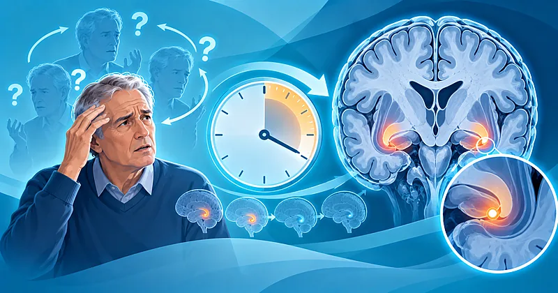
Transient global amnesia is a sudden, dense anterograde amnesia in an otherwise intact adult that resolves within 24 hours and carries an excellent prognosis. What has changed is not the syndrome but how confidently it can be diagnosed, when imaging actually helps, and what patients can be told about the future. The points below should reshape the next emergency assessment of a transient global amnesia episode.
Key Clinical Takeaways
Each action below is expanded in the section it links to, with thresholds and evidence tags.
- 1Diagnose transient global amnesia at the bedside when a witnessed, isolated anterograde amnesia with repetitive questioning occurs without focal signs, altered consciousness, or loss of personal identity → Is This Transient Global Amnesia?
- 2Treat any focal deficit, headache, vomiting, fever, drowsiness, or amnesia beyond 24 hours as a red flag requiring urgent imaging and a mimic-directed work-up → Which Features Mean It Is Not TGA?
- 3Check capillary glucose, basic bloods, and an ECG in every patient, and reserve urgent imaging for atypical features, trauma, or anticoagulation → Which Tests Does a Typical Episode Need?
- 4Time confirmatory MRI to about 24–72 hours after onset with thin-slice DWI through the hippocampus, and do not treat an early negative scan as excluding the diagnosis → When and How Should MRI Be Done?
- 5Order an EEG when attacks are brief, recurrent, occur on waking, or number more than three in a year → When Should EEG Be Ordered?
- 6Observe until new memories are forming, and discharge only to a responsible adult with clear return advice → Admit, Observe, or Discharge?
- 7Counsel that stroke risk is not increased, recurrence is uncommon, and the dementia signal is uncertain, while treating vascular risk factors as usual → What Should I Tell the Patient?
Why This Update Matters
Transient global amnesia has an estimated incidence of 3–8 per 100,000 per year, and about three-quarters of patients are aged 50–70 years.1 Its presentation is dramatic, so it generates emergency attendances, imaging, and admissions out of proportion to its risk. At the same time, a posterior circulation stroke, encephalitis, or a focal seizure can look similar for the first hour. Getting the diagnosis right saves unnecessary investigation, avoids inappropriate antithrombotic therapy, and protects the minority whose amnesia signals something treatable.
Is This Transient Global Amnesia? Making a Positive Bedside Diagnosis
The patient is alert, communicative, and able to perform complex, well-learned tasks, yet cannot retain new information for more than a minute or two and asks the same questions repeatedly.1,15 The new-learning span shrinks to roughly 30–180 seconds, orientation to time and place is lost, and orientation to self is always preserved.1 A variable retrograde amnesia for hours to days is common, and after recovery a permanent gap covering the attack remains.
Episodes last on average 6–8 hours and by definition resolve within 24 hours; a typical episode shorter than an hour does not argue against transient global amnesia.1,23 Up to 85% follow a precipitant such as physical exertion, a Valsalva-like activity, cold-water immersion, sexual intercourse, or emotional stress.1,22
Bedside Criteria Translated Into Checks
The classic Caplan and Hodges–Warlow criteria are reorganised below into what to check at the bedside and what would overturn the diagnosis.1,2
| Criterion | How to Check It at the Bedside | What Breaks the Diagnosis |
|---|---|---|
| Witnessed attack | Take the history from the companion, not only the patient | No observer and no corroboration (consider MRI support) |
| Clear anterograde amnesia | Three-word recall at 5 minutes; repeated questions | Patient recounts details and timing of the episode |
| Consciousness and identity intact | Knows own name; fully alert | Drowsiness, confusion beyond memory, loss of identity |
| Cognition otherwise intact | Naming, language, praxis, and attention preserved | Aphasia, neglect, apraxia, or visual field loss |
| No focal neurological signs | Full examination, including visual fields and gait | Any focal deficit |
| Resolution within 24 hours | Serial memory testing until new learning returns | Persistent deficit beyond 24 hours |
| No epilepsy or recent head injury | Seizure history, antiseizure drugs, trauma | Active epilepsy or head injury |
Diagnose transient global amnesia clinically using established criteria, obtaining the history from a reliable witness. Practice impact: make the diagnosis positively in the emergency department rather than by exhaustive exclusion.
Strong RecModerate EvidenceDGN 2023Hodges & Warlow 1990Test anterograde memory formally with delayed word-list recall, and repeat it until new learning returns, documenting the time of recovery.
Moderate RecLow EvidenceDGN 2023Perform a complete neurological examination, including visual fields and language, before accepting the label.
Strong RecLow EvidenceDGN 2023Ask specifically about the preceding hours — exertion, immersion, intercourse, emotional stress, or a procedure — because an identifiable precipitant supports the diagnosis.
Moderate RecLow EvidenceQuinette 2006DGN 2023Consider the diagnosis in typical episodes lasting under an hour, rather than dismissing it on duration alone.
Conditional RecLow EvidenceRomoli 2020Which Features Mean It Is Not TGA? Red Flags and Mimics
Anything beyond isolated amnesia with mild autonomic symptoms argues against transient global amnesia: drowsiness, severe headache, vomiting, fever, confusion, focal signs, isolated retrograde amnesia, or failure to recover by 24 hours.1 Age under 50 is itself a warning, because the syndrome is rare in that group and has not been described below 30.1
| Mimic | Clue That Separates It | First Discriminating Step |
|---|---|---|
| Posterior cerebral artery or hippocampal infarct | Visual field defect, sensory signs, persistent deficit, vascular risk | Urgent MRI with DWI and vessel imaging |
| Transient epileptic amnesia | Brief (often under an hour), recurrent, on waking, autobiographical memory loss | EEG, ideally with sleep; seizure history |
| Viral or autoimmune encephalitis | Fever, headache, seizures, behavioural change, persistent deficit | MRI, CSF, and early empirical aciclovir if suspected |
| Hypoglycaemia or metabolic encephalopathy | Diabetes treatment, sweating, fluctuating consciousness | Capillary glucose, electrolytes |
| Drug or alcohol amnesia | Benzodiazepines, Z-drugs, alcohol, anticholinergics | Medication and intake history; toxicology if unclear |
| Post-traumatic amnesia | Fall or head strike, anticoagulation | CT head |
| Functional or dissociative amnesia | Loss of personal identity; inconsistent pattern | Collateral history; psychiatric assessment |
Image urgently, preferably with MRI, when the presentation is atypical or any red flag is present. Practice impact: atypical features move the patient from reassurance to a time-critical stroke or encephalitis pathway.
Strong RecLow EvidenceDGN 2023Evaluate rapidly for alternative causes in any patient under 50 years presenting with suspected transient global amnesia.
Strong RecLow EvidenceDGN 2023Exclude posterior circulation stroke with MRI whenever amnesia is accompanied by a focal deficit, including a subtle hemianopia.
Strong RecModerate EvidenceDGN 2023Arena 2015Start the encephalitis pathway, including empirical aciclovir where appropriate, when amnesia is accompanied by fever, headache, seizures, or persistent confusion.
Strong RecLow EvidenceDGN 2023Avoid labelling an unwitnessed episode as transient global amnesia on history alone; seek collateral information or imaging support.
Moderate RecLow EvidenceSzabo 2020Which Tests Does a Typical Episode Need?
For a witnessed, typical episode that is resolving, very little testing is required.1,3 The aim is to exclude common, dangerous mimics cheaply — hypoglycaemia, metabolic derangement, and cardiac events — rather than to prove transient global amnesia. Asymptomatic troponin elevation has been reported in a minority of patients, and a link with stress cardiomyopathy has been described, so the ECG earns its place.1,20
Check capillary glucose immediately in every patient presenting with acute amnesia.
Strong RecLow EvidenceDGN 2023Obtain basic bloods (electrolytes, renal function, full blood count) and a 12-lead ECG in all patients.
Moderate RecLow EvidenceArena 2015Consider troponin when there is chest discomfort, ECG change, or recent intense emotional or physical stress.
Conditional RecLow EvidenceDGN 2023Erdur 2019Perform CT when head injury or anticoagulation is present and MRI is not promptly available, because trauma excludes the diagnosis and haemorrhage must be ruled out.
Strong RecLow EvidenceDGN 2023Avoid routine emergency MRI in typical, resolving episodes seen within the first hours, where the yield for confirmation is low. Practice impact: stop ordering reflexive stroke-protocol MRI for classic, resolving episodes.
Moderate RecModerate EvidenceWong 2022DGN 2023Do not order SPECT or PET for diagnosis.
AgainstLow EvidenceDGN 2023When and How Should MRI Be Done to Confirm Transient Global Amnesia?
The imaging signature is one or more punctate, 1–5 mm foci of restricted diffusion in the hippocampus, typically in the CA1 region, which later show T2 signal and then disappear without a lasting lesion.1,16 In a series of 390 consecutive patients, hippocampal DWI lesions supported the diagnosis in 70.6%, including 69.1% of those with lower clinical certainty, and lesions were visible up to 6 days after onset.4
Timing drives yield. A systematic review of 23 studies found positivity of about 15.6% in the first 12 hours, rising to roughly 66–83% in later windows, with wide confidence intervals.5 A separate meta-analysis estimated overall yield at 39%, rising to 63% with slices of 3 mm or less and up to 68% at more than 24 to 96 hours.6
A Hippocampus-Focused Protocol
Radiology departments often default to a stroke protocol, which uses thicker slices and may miss the lesion. The settings below are guideline-described; confirm them with local neuroradiology.1,17
| Parameter | Recommended Setting | Why It Matters |
|---|---|---|
| Timing | About 24–72 hours after onset | Lesions become most conspicuous after the first day |
| Field strength | 3 T where available | Higher detection of small lesions |
| DWI slice thickness | 3 mm or less | Reduces partial-volume loss of tiny foci |
| T2 slice thickness | About 2 mm | Captures evolving T2 signal |
| b-value | High (2000–3000 s/mm²) | Improves lesion conspicuity |
| Orientation | Axial and coronal along the hippocampal long axis | Brings the CA1 region into plane |
| Sequences | DWI, ADC, T2 | Confirms true restriction and excludes other pathology |
Schedule confirmatory MRI about 24–72 hours after onset when the diagnosis needs support, rather than in the first hours. Practice impact: book next-day outpatient or ward MRI instead of an immediate scan for a typical case.
Moderate RecModerate EvidenceDGN 2023Szabo 2020Wong 2022Request thin-slice (≤3 mm) DWI with coronal views through the hippocampi, and state the clinical question on the request.
Moderate RecModerate EvidenceLim 2021Weon 2008Use MRI particularly when clinical certainty is low — an unwitnessed episode, a self-reported amnestic gap, or incomplete history.
Moderate RecModerate EvidenceSzabo 2020Do not exclude transient global amnesia because DWI is negative, especially if the scan was early or used thick slices.
Strong RecModerate EvidenceDGN 2023Wong 2022Perform vascular imaging and a cardiac embolic source work-up when DWI lesions appear outside the hippocampus.
Moderate RecLow EvidenceDGN 2023Ganeshan 2022Clinical Decision Pathway
A question-based route from first contact to discharge for suspected transient global amnesia.
Admit, Observe, or Discharge?
Most patients can go home once the amnesia has resolved. Admission adds little for a typical, resolved episode but is appropriate when the diagnosis is in doubt or when nobody can supervise a still-amnestic patient.1
Observe the patient until anterograde memory has recovered, confirmed by delayed recall of new information.
Moderate RecLow EvidenceDGN 2023Consider inpatient monitoring for up to 24 hours or until symptoms resolve when no relative or carer can supervise the patient.
Conditional RecLow EvidenceDGN 2023Admit patients with persistent symptoms or features that cast doubt on the diagnosis, for imaging and observation.
Strong RecLow EvidenceDGN 2023Discharge patients whose episode has fully resolved and fits the criteria, without routine admission. Practice impact: a resolved, typical episode is a discharge, not an admission for “TIA work-up”.
Moderate RecLow EvidenceDGN 2023Arena 2015Give written information to both patient and companion, because the patient will not remember the verbal explanation given during the attack.
Moderate RecLow EvidenceArena 2015When Should EEG Be Ordered, and Could It Be Epilepsy?
Transient epileptic amnesia is the most important recurrent mimic. It presents in middle to late life with brief, recurrent amnestic episodes, often on waking, and is associated with accelerated forgetting between attacks, patchy loss of autobiographical memories, and a good response to antiseizure medication.12 In early follow-up studies, about 7% of patients labelled with transient global amnesia later developed epilepsy, mostly those with brief or recurrent attacks.2
| Feature | Transient Global Amnesia | Transient Epileptic Amnesia |
|---|---|---|
| Typical duration | Several hours (average 6–8) | Usually under an hour |
| Frequency | Usually single; recurrence uncommon | Recurrent, often several per year |
| Timing | Often after a precipitant, while awake | Frequently on waking |
| Repetitive questioning | Characteristic | May occur |
| Memory between attacks | Normal | Accelerated forgetting, autobiographical gaps |
| EEG | Normal or non-specific | May show temporal epileptiform discharges; can be normal |
| Response to antiseizure drugs | Not applicable | Usually prompt |
Order an EEG when amnestic attacks are brief, recurrent, occur on waking, or number more than three per year. Practice impact: frequent short episodes are an epilepsy work-up, not repeated reassurance.
Moderate RecLow EvidenceDGN 2023Arena 2015Consider sleep-deprived or prolonged EEG when the standard recording is normal but suspicion of transient epileptic amnesia remains, because a normal EEG does not exclude it.
Conditional RecLow EvidenceDGN 2023Butler 2007Refer to an epilepsy service when attacks are recurrent and stereotyped, particularly with persistent interictal memory complaints.
Moderate RecLow EvidenceButler 2007Avoid routine EEG after a single, typical, witnessed episode.
Conditional RecLow EvidenceArena 2015Who Needs a Different Approach?
Several groups need the transient global amnesia framework adjusted rather than abandoned.
Investigate adults under 50 years comprehensively, because transient global amnesia is rare at that age and alternative diagnoses are more likely.
Strong RecLow EvidenceDGN 2023Support the diagnosis with delayed thin-slice MRI when the episode was unwitnessed or the history comes only from the patient.
Moderate RecModerate EvidenceSzabo 2020Evaluate patients with recurrent episodes for transient epileptic amnesia and for migraine, which is over-represented among recurrences.
Moderate RecModerate EvidenceMorris 2020Hernández 2022Assess cardiac status in patients whose episode followed intense emotional stress or who have chest symptoms, given reported myocardial injury.
Conditional RecLow EvidenceErdur 2019Consider an amnestic presentation of another disorder in patients with existing cognitive impairment, where baseline memory confounds the history.
Conditional RecLow EvidenceArena 2015What Should I Tell the Patient About Transient Global Amnesia?
Patients and families ask three questions: will it happen again, was it a stroke, and is it the start of dementia? The evidence supports clear, mostly reassuring answers, with one honest caveat.
| The Question Patients Ask | What the Evidence Shows | A Useful Way to Say It |
|---|---|---|
| “Will it come back?” | About 12.7% recurrence in a meta-analysis of 4,514 cases; most who recur have three or fewer episodes | “Most people have only one episode; about one in eight has another.” |
| “Who is more likely to have another?” | Migraine (OR ~2.1), depression (OR ~4.5), sex as trigger (OR ~1.5), younger first episode | “Your migraine history slightly raises the chance of a repeat.” |
| “Was it a stroke? Will I have one?” | Annual stroke risk 0.6% and 5-year 2.7% in 525 patients, no higher than expected | “This was not a stroke, and it does not raise your stroke risk.” |
| “Has it damaged my memory?” | Subtle test differences for days to months; no lasting impairment in clinical cohorts | “You may feel off for a few days; memory recovers.” |
| “Does it lead to dementia?” | No excess in a 12-year matched cohort; a Korean claims cohort reported an adjusted HR of 1.40 | “Most studies show no link; one large database study found a small association that is still being examined.” |
Recurrence data come from a meta-analysis and a 1,044-patient Mayo Clinic cohort in which 13.7% had recurrent episodes and 95.8% of these had three or fewer.10,11 In a population-based cohort of 221 patients followed for a mean of 12 years, there was no excess of cerebrovascular events, seizures, or cognitive impairment compared with matched controls.9 In two prospective cohorts totalling 525 patients, stroke risk was no greater than expected for the population, and administrative and propensity-matched analyses agree.8,18,19 Subtle neuropsychological differences can be detected for days to months after an episode but have not been shown to persist.1,21
Reassure patients that transient global amnesia does not increase stroke risk, and do not start antiplatelet therapy for the episode itself. Practice impact: remove “TIA” from the discharge letter and avoid reflexive aspirin.
Moderate RecModerate EvidenceRomoli 2019Arena 2017Garg 2021Identify and treat cardiovascular risk factors according to usual primary-prevention guidance.
Moderate RecLow EvidenceDGN 2023Counsel that roughly one in eight patients has a recurrence, higher with migraine, depression, or a younger first episode.
Moderate RecModerate EvidenceHernández 2022Morris 2020Do not prescribe prophylactic medication to prevent recurrence, as no treatment has evidence of benefit.
AgainstLow EvidenceDGN 2023Advise that transient symptoms such as fatigue or feeling “not quite right” can persist for several days, and that formal testing may show subtle, temporary changes.
Moderate RecLow EvidenceSandikci 2022DGN 2023Discuss the dementia literature honestly when asked, framing the recent association as unconfirmed and confounded, and offer review if new cognitive symptoms appear.
Conditional RecLow EvidenceLee 2024Liampas 2021Advise patients to seek urgent care if a future episode includes weakness, speech or vision change, severe headache, or lasts beyond 24 hours.
Moderate RecLow EvidenceDGN 2023Monitoring and Follow-Up
Long-term follow-up is not required after a single, typical episode. Review is aimed at the patients whose course departs from the expected pattern.
Arrange neurology review for recurrent episodes, atypical features, extrahippocampal DWI lesions, or persistent memory complaints beyond a few weeks.
Moderate RecLow EvidenceDGN 2023Reassess the diagnosis if a second episode is brief, occurs on waking, or is followed by interictal memory complaints.
Moderate RecLow EvidenceButler 2007Consider formal neuropsychological assessment when memory complaints persist beyond the expected recovery period.
Conditional RecLow EvidenceDGN 2023Document the episode as transient global amnesia in the primary-care record, including any confirmatory MRI, to prevent future mislabelling.
Moderate RecLow EvidenceArena 2015| What to Watch | When | What Triggers Action | Action |
|---|---|---|---|
| Anterograde memory | During the episode, until recovery | No return of new learning by 24 hours | Full work-up for a mimic |
| New neurological symptoms | During observation and after discharge | Weakness, speech, vision, headache, seizure | Emergency reassessment and imaging |
| Recurrence | Any future episode | Brief, frequent, or waking episodes | EEG and epilepsy referral |
| Memory complaints | Weeks to months | Persistence beyond the expected recovery | Neuropsychology and neurology review |
| Vascular risk factors | Routine primary care | Uncontrolled blood pressure, lipids, diabetes | Usual primary-prevention management |
Evidence in Context
Where the transient global amnesia evidence converges, where it diverges, and what newer studies add.
Where Sources Agree▾
Guidelines and major reviews agree that transient global amnesia is a clinical diagnosis, that typical resolving episodes need minimal testing, that urgent imaging is for atypical presentations, and that EEG is reserved for brief or recurrent attacks.
Where They Differ on MRI▾
The German guideline and imaging series treat delayed DWI as a positive confirmatory test, while an emergency-medicine meta-analysis emphasises its low early yield and questions routine acute scanning. The positions reconcile when MRI is timed for confirmation rather than used as an emergency screen.
Mechanism Remains Uncertain▾
Venous congestion after Valsalva manoeuvres, migraine-like spreading depression, stress-related hippocampal vulnerability, and arterial ischaemia have all been proposed. The absence of excess stroke risk argues against arterial ischaemia as the usual cause.
The Dementia Question▾
Clinical cohorts with careful case ascertainment found no excess cognitive decline, but Taiwanese and Korean administrative databases report associations. Coding-based diagnoses may include misclassified early neurodegeneration or epilepsy, and residual confounding is likely, so the signal needs prospective confirmation.
Evidence Gaps
These are the open questions a clinician actually meets with transient global amnesia, where practice rests on observational data and consensus.
- Whether transient global amnesia is causally linked to later dementia, or whether administrative-cohort associations reflect misclassification and confounding, is unresolved.
- No trial has tested any intervention to prevent recurrence, including in patients with migraine.
- The optimal MRI timing and protocol come from heterogeneous observational series; the diagnostic accuracy of the clinical criteria themselves has not been formally validated.
- How often extrahippocampal DWI lesions represent true embolic events, and whether they change management, is uncertain.
- Evidence-based driving and occupational advice after a single or recurrent episode does not exist.
References
- 1.Sander D, Bartsch T, Connolly F, et al. Guideline “Transient Global Amnesia (TGA)” of the German Society of Neurology (Deutsche Gesellschaft für Neurologie): S1-guideline. Neurol Res Pract. 2023;5:15. https://doi.org/10.1186/s42466-023-00240-0
- 2.Hodges JR, Warlow CP. Syndromes of transient amnesia: towards a classification. A study of 153 cases. J Neurol Neurosurg Psychiatry. 1990;53(10):834-843. https://doi.org/10.1136/jnnp.53.10.834
- 3.Arena JE, Rabinstein AA. Transient global amnesia. Mayo Clin Proc. 2015;90(2):264-272. https://doi.org/10.1016/j.mayocp.2014.12.001
- 4.Szabo K, Hoyer C, Caplan LR, et al. Diffusion-weighted MRI in transient global amnesia and its diagnostic implications. Neurology. 2020;95(2):e206-e212. https://doi.org/10.1212/WNL.0000000000009783
- 5.Wong ML, Silva LOJ, Gerberi DJ, Edlow JA, Dubosh NM. Sensitivity of diffusion-weighted magnetic resonance imaging in transient global amnesia as a function of time from symptom onset. Acad Emerg Med. 2022;29(4):398-405. https://doi.org/10.1111/acem.14390
- 6.Lim SJ, Kim M, Suh CH, et al. Diagnostic yield of diffusion-weighted brain magnetic resonance imaging in patients with transient global amnesia: a systematic review and meta-analysis. Korean J Radiol. 2021;22(10):1680-1689. https://pubmed.ncbi.nlm.nih.gov/34269537/
- 7.Ganeshan R, Betz M, Scheitz JF, et al. Frequency of silent brain infarction in transient global amnesia. J Neurol. 2022;269(3):1422-1426. https://doi.org/10.1007/s00415-021-10705-4
- 8.Romoli M, Tuna MA, McGurgan I, et al. Long-term risk of stroke after transient global amnesia in two prospective cohorts. Stroke. 2019;50(9):2555-2557. https://doi.org/10.1161/STROKEAHA.119.025720
- 9.Arena JE, Brown RD, Mandrekar J, Rabinstein AA. Long-term outcome in patients with transient global amnesia: a population-based study. Mayo Clin Proc. 2017;92(3):399-405. https://doi.org/10.1016/j.mayocp.2016.11.015
- 10.Hernández MA, Arena JE, Alessandro L, Allegri RF, Calandri IL. Transient global amnesia recurrence: prevalence and risk factor meta-analysis. Neurol Clin Pract. 2022;12(4):e35-e48. https://doi.org/10.1212/CPJ.0000000000001181
- 11.Morris KA, Rabinstein AA, Young NP. Factors associated with risk of recurrent transient global amnesia. JAMA Neurol. 2020;77(12):1551-1558. https://doi.org/10.1001/jamaneurol.2020.2943
- 12.Butler CR, Graham KS, Hodges JR, et al. The syndrome of transient epileptic amnesia. Ann Neurol. 2007;61(6):587-598. https://doi.org/10.1002/ana.21111
- 13.Lee SJ, Lee TK, Bae YJ, Kim M. Increased risk of dementia after transient global amnesia: a nationwide population-based, longitudinal follow-up study in South Korea. Neuroepidemiology. 2024;58(4):247-255. https://pubmed.ncbi.nlm.nih.gov/38295784/
- 14.Liampas I, Raptopoulou M, Siokas V, et al. The long-term prognosis of transient global amnesia: a systematic review. Rev Neurosci. 2021;32(5):531-543. https://doi.org/10.1515/revneuro-2020-0110
- 15.Ropper AH. Transient global amnesia. N Engl J Med. 2023;388(7):635-640. https://doi.org/10.1056/NEJMra2213867
- 16.Bartsch T, Deuschl G. Transient global amnesia: functional anatomy and clinical implications. Lancet Neurol. 2010;9(2):205-214. https://doi.org/10.1016/S1474-4422(09)70344-8
- 17.Weon YC, Kim JH, Lee JS, Kim SY. Optimal diffusion-weighted imaging protocol for lesion detection in transient global amnesia. AJNR Am J Neuroradiol. 2008;29(7):1324-1328. https://doi.org/10.3174/ajnr.A1105
- 18.Mangla A, Navi BB, Layton K, Kamel H. Transient global amnesia and the risk of ischemic stroke. Stroke. 2014;45(2):389-393. https://doi.org/10.1161/STROKEAHA.113.003916
- 19.Garg A, Limaye K, Shaban A, Adams HP, Leira EC. Transient global amnesia does not increase the risk of subsequent ischemic stroke: a propensity score-matched analysis. J Neurol. 2021;268(9):3301-3306. https://doi.org/10.1007/s00415-021-10483-z
- 20.Erdur H, Siegerink B, Ganeshan R, et al. Myocardial injury in transient global amnesia: a case-control study. Eur J Neurol. 2019;26:986-991. https://doi.org/10.1111/ene.13920
- 21.Sandikci V, Ebert A, Zurwesten L, et al. The remains of the day: neuropsychological findings in postacute transient global amnesia. J Neurol. 2022;269(9):4764-4771. https://doi.org/10.1007/s00415-022-11110-1
- 22.Quinette P, Guillery-Girard B, Dayan J, et al. What does transient global amnesia really mean? Review of the literature and thorough study of 142 cases. Brain. 2006;129(7):1640-1658. https://doi.org/10.1093/brain/awl105
- 23.Romoli M, Tuna MA, Li L, et al. Time trends, frequency, characteristics and prognosis of short-duration transient global amnesia. Eur J Neurol. 2020;27(5):887-893. https://doi.org/10.1111/ene.14163
How to Read the Evidence Tags
Every recommendation carries a strength tag and an evidence tag — Medaptly’s own simplified reading of the underlying guidance, not a reproduction of any source body’s grading system.
| Strength Tag | What It Means |
|---|---|
| Strong Rec | High-quality evidence broadly supports this action |
| Moderate Rec | The weight of evidence favours this action |
| Conditional Rec | The benefit is less certain — individualise |
| Against | Evidence points to no benefit or potential harm |
| Evidence Tag | What It Means |
|---|---|
| High Evidence | Multiple well-designed trials or high-quality meta-analyses |
| Moderate Evidence | A single trial or large observational dataset |
| Low Evidence | Expert consensus or small studies |