
Vulvar Cancer Treatment: 8 Essential Pathway Decisions
Clinical Practice Update — Biopsy Site Selection, Sentinel Lymph Node Mapping, and Surgical Margins
This is an original clinical education article informed by current guidelines and evidence. See References below for source documents.
- Clinical Focus
- Evidence-based vulvar cancer treatment with emphasis on biopsy technique, groin staging, and resection margins
- Target Audience
- Gynaecologic oncologists, general gynaecologists, surgical trainees, primary care physicians screening symptomatic women
- Setting
- Outpatient gynaecology clinic, vulvar dermatology, gynaecologic oncology operating theatre
- Source Evidence
- •NCCN Clinical Practice Guidelines in Oncology — Vulvar Cancer (Squamous Cell Carcinoma), Version 2.2024
- •ESGO-ESTRO-ESP Guidelines for the Management of Patients with Vulvar Cancer — Update 2023
- •GROINSS-V I (Van der Zee, JCO 2008) and GROINSS-V II (Oonk, JCO 2021)
- •RCOG Green-top Guideline No. 33 — Management of Vulval Cancer
Key Clinical Takeaways
Effective vulvar cancer treatment in 2026 rests on three pivots: precise biopsy site selection at first presentation, accurate groin staging through sentinel lymph node mapping, and oncologically adequate yet conservative surgical margins. The points below condense the modern pathway into eight rules clinicians can apply from the first abnormal vulvar exam through definitive surgery.
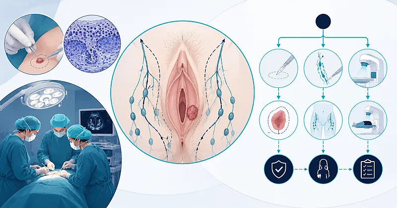
- 1Take a punch biopsy from the edge of the most suspicious area, capturing the transition between abnormal and adjacent skin → Biopsy Approach
- 2Use mapping biopsies for heterogeneous or multifocal disease — one biopsy from one site rarely captures the full picture → Biopsy Approach
- 3Offer sentinel lymph node biopsy for unifocal tumours under 4 cm with clinically and radiologically negative groins → Sentinel Lymph Node
- 4Use combined technique (radiocolloid plus blue dye) for sentinel node identification — the dual approach maximises detection → Sentinel Lymph Node
- 5Map bilaterally for any tumour within 1 cm of the midline; unilateral mapping is enough only for clearly lateralised lesions → Sentinel Lymph Node
- 6Aim for a clinical (gross) excision margin of at least 1 cm at surgery, but accept narrower pathologic margins when no other risk factors are present → Surgical Margins
- 7For sentinel node micrometastases of 2 mm or smaller, adjuvant inguinofemoral radiotherapy is a proven alternative to completion lymphadenectomy (GROINSS-V II) → Adjuvant Therapy
- 8Centralise care — long-term outcomes of vulvar cancer treatment are best in specialist gynaecologic oncology centres → Where to Treat
Biopsy Approach in Vulvar Cancer Treatment
A diagnostic biopsy that captures the full picture of invasion is the single most important first step in vulvar cancer treatment. The pathology report from this biopsy drives every later decision: the FIGO stage, the need for groin staging, the planned excision margin, and the choice between surgery and primary chemoradiation in advanced disease.
Vulvar lesions often present in the context of vulvar intraepithelial neoplasia or chronic dermatoses, where the surface change can be patchy and the area of true invasion small. A biopsy taken from the wrong site — or too superficially — can miss invasion entirely, delay the cancer diagnosis, and trigger an inadequate first surgery.
In women with longstanding lichen sclerosus or differentiated vulvar intraepithelial neoplasia, any new ulcer, indurated plaque, or non-healing lesion that has not regressed on topical steroids within 6 to 8 weeks should be biopsied. The threshold for biopsy in this population is low.
Recommendations
Perform a Keyes punch biopsy (4–6 mm) from the edge of the suspicious lesion, capturing both the abnormal area and 1–2 mm of clinically uninvolved skin to assess invasion depth and the transition zone.
Strong Rec High Evidence NCCN 2024 ESGO 2023Take multiple mapping biopsies for large (>2 cm), multifocal, or heterogeneous lesions — one biopsy per clinically distinct area — to avoid missing invasive disease in a field of intraepithelial change.
Strong Rec Moderate Evidence ESGO 2023Avoid shave biopsy when invasive disease is suspected — a shave specimen often fails to capture depth and forces a repeat procedure before vulvar cancer treatment can proceed.
Moderate Rec Moderate Evidence NCCN 2024Do not biopsy from the centre of a necrotic or heavily ulcerated lesion — the resulting sample is usually non-diagnostic and forces a return visit.
Against Low Evidence Expert ConsensusDocument each biopsy site with a clinical photograph and an annotated diagram of the vulva. Precise localisation matters when a small lesion is no longer visible after excision biopsy.
Strong Rec Low Evidence ESGO 2023Specifically request that pathology report depth of invasion in millimetres, horizontal tumour size, lymphovascular space invasion, and the histologic subtype — not just “invasive carcinoma present.”
Strong Rec High Evidence NCCN 2024 ESGO 2023Staging and Imaging Before Surgery
Vulvar cancer is surgically staged using the FIGO 2021 system. Depth of invasion, tumour size, groin node status, and distant spread together drive whether vulvar cancer treatment is primarily surgical, primarily chemoradiation, or a combination.
Stage IA disease (lesion ≤2 cm with depth of invasion ≤1 mm) carries a node metastasis risk under 1% and does not require groin assessment. Once invasion exceeds 1 mm or the tumour is larger than 2 cm, groin staging becomes mandatory because nodal status is the strongest single predictor of survival.
Recommendations
Obtain pelvic MRI for any tumour >4 cm, suspected sphincter or urethral involvement, or clinically palpable groin nodes — MRI is the best modality for local extent.
Strong Rec Moderate Evidence NCCN 2024 ESGO 2023Use groin ultrasound with fine-needle aspiration of any sonographically suspicious node — a positive FNA spares the patient an unnecessary sentinel node procedure.
Strong Rec Moderate Evidence ESGO 2023Consider FDG-PET/CT in locally advanced disease (T3/T4) or where MRI suggests extra-pelvic spread, particularly when concurrent chemoradiation is being planned.
Moderate Rec Moderate Evidence NCCN 2024Test HPV/p16 status on the diagnostic biopsy — HPV-associated disease carries a more favourable prognosis and the result is now reported in modern pathologic staging.
Moderate Rec Moderate Evidence ESGO 2023Sentinel Lymph Node in Vulvar Cancer Treatment
Sentinel lymph node biopsy is the most important morbidity-reducing innovation in vulvar cancer treatment of the last two decades. The GROINSS-V I study demonstrated that for early-stage, unifocal, clinically node-negative tumours, a negative sentinel node carries a groin recurrence rate around 3% and survival comparable to full inguinofemoral lymphadenectomy — with markedly less lymphoedema, wound breakdown, and cellulitis.
The decision between sentinel node mapping and inguinofemoral lymphadenectomy hinges on three tumour features: size, focality, and clinical groin status. Outside the strict GROINSS-V eligibility criteria, completion lymphadenectomy remains standard.
Patient Selection
All five must apply: squamous histology, unifocal tumour, maximum diameter <4 cm, depth of invasion >1 mm, and clinically and radiologically negative groin nodes. Failure of any one shifts the patient to inguinofemoral lymphadenectomy.
Recommendations
Offer sentinel lymph node biopsy as the preferred groin staging procedure for women with unifocal squamous vulvar carcinoma <4 cm with clinically and radiologically negative groins.
Strong Rec High Evidence GROINSS-V I NCCN 2024 ESGO 2023Use the combined technique — preoperative technetium-99m radiocolloid lymphoscintigraphy plus intraoperative blue dye — for sentinel node identification. Combined tracing maximises detection compared with either technique alone.
Strong Rec High Evidence GROINSS-V I ESGO 2023Perform bilateral sentinel node mapping for any tumour located within 1 cm of the midline. Lateralised tumours (>1 cm from midline) may have unilateral mapping, with completion contralateral assessment only if the ipsilateral side is positive.
Strong Rec Moderate Evidence NCCN 2024 ESGO 2023If no sentinel node is identified intraoperatively despite combined tracer use, proceed to ipsilateral inguinofemoral lymphadenectomy at the same operation — do not leave the groin unstaged.
Strong Rec Moderate Evidence NCCN 2024 ESGO 2023Send sentinel nodes for ultrastaging with serial sectioning at 200—400 micron intervals and immunohistochemistry for cytokeratins — standard single-section histology misses up to 23% of micrometastases.
Strong Rec High Evidence GROINSS-V I ESGO 2023Do not offer sentinel lymph node biopsy for tumours ≥4 cm, multifocal disease, or any palpable or sonographically suspicious groin node — the false-negative rate becomes unacceptable.
Against Moderate Evidence NCCN 2024 ESGO 2023Surgical Margins for Effective Vulvar Cancer Treatment
Few topics in vulvar cancer treatment have shifted as much in the last decade as the optimal surgical margin. The traditional teaching — that a tumour-free pathologic margin of less than 8 mm predicts local recurrence — was based on a small 1990s series and has been challenged by multiple modern cohorts. Current practice balances oncologic clearance against the functional and psychosexual consequences of overly radical resection.
Two concepts are now distinguished. The gross (clinical) margin is what the surgeon measures with a ruler at the time of excision and is targeted at ≥1 cm to allow for tissue retraction. The pathologic margin is what the pathologist measures on the fixed specimen and is usually 25–30% smaller because tissue contracts after fixation. The 1 cm clinical margin therefore typically yields a 7–8 mm pathologic margin.
Recommendations
At surgery, aim for a clinical (gross) excision margin of at least 1 cm circumferentially, allowing for fixation shrinkage to give a pathologic margin in the 7–8 mm range.
Strong Rec Moderate Evidence NCCN 2024 ESGO 2023Accept a pathologic tumour-free margin between 3 mm and 8 mm without automatic re-excision, provided no other adverse features are present (no lymphovascular invasion, no perineural invasion, no high-grade histology, node-negative).
Conditional Rec Moderate Evidence ESGO 2023 Woelber 2016Re-excise to clear margins when the pathologic tumour-free margin is <3 mm or when any margin is positive (R1/R2), provided functional re-resection is feasible.
Strong Rec Moderate Evidence NCCN 2024 ESGO 2023Offer adjuvant radiotherapy to the vulva when re-excision is not feasible due to anatomic constraints (sphincter, urethra, clitoris) or when the patient declines further surgery.
Moderate Rec Moderate Evidence NCCN 2024Prefer wide local excision (also called radical local excision) over historic radical vulvectomy for unifocal tumours. Long-term recurrence and survival are equivalent, with substantially less morbidity.
Strong Rec High Evidence NCCN 2024 ESGO 2023Tailor the deep margin to the underlying perineal membrane — resection should reach the inferior fascia of the urogenital diaphragm. Compromise of the deep margin matters more for local recurrence than a narrow peripheral margin.
Moderate Rec Moderate Evidence ESGO 2023Adjuvant Therapy After Surgery
Adjuvant treatment after vulvar cancer treatment is driven mainly by lymph node status. The GROINSS-V II trial reshaped practice for sentinel node micrometastases ≤2 mm, showing that adjuvant inguinofemoral radiotherapy gives groin recurrence rates comparable to completion lymphadenectomy with far less morbidity.
For macrometastases (>2 mm), completion inguinofemoral lymphadenectomy followed by adjuvant radiotherapy remains the standard. The use of concurrent chemotherapy — typically weekly cisplatin — is extrapolated from cervical cancer practice and is offered for extracapsular extension or multiple positive nodes.
For sentinel node micrometastases ≤2 mm, offer adjuvant inguinofemoral radiotherapy (50 Gy) as an alternative to completion lymphadenectomy.
Moderate Rec High Evidence GROINSS-V II ESGO 2023For sentinel node macrometastases (>2 mm), proceed to completion inguinofemoral lymphadenectomy followed by adjuvant groin and pelvic radiotherapy.
Strong Rec High Evidence NCCN 2024 GROINSS-V IIConsider concurrent weekly cisplatin (40 mg/m²) with adjuvant radiotherapy when there is extracapsular nodal extension or two or more positive nodes.
Moderate Rec Moderate Evidence NCCN 2024For locally advanced unresectable disease (T3/T4), offer primary chemoradiation rather than ultraradical surgery — outcomes are comparable and quality of life is better preserved.
Strong Rec Moderate Evidence NCCN 2024Clinical Decision Pathway
A question-based walk-through of vulvar cancer treatment from suspected lesion to definitive plan. Use each question in order; the answer to one determines whether the next applies.
Practical Tables for Bedside Use
Biopsy Technique by Lesion Characteristic
| Lesion Type | Preferred Technique | Specimen Yield Target | Practical Tip |
|---|---|---|---|
| Discrete plaque or ulcer <2 cm | Keyes punch 4–6 mm | Full-thickness skin + 2–3 mm dermis | Sample the edge, not the centre |
| Heterogeneous area >2 cm | Multiple mapping punch biopsies | One per clinically distinct area | Label each site on a vulvar diagram |
| Suspected microinvasion, <2 cm | Excisional biopsy, 5 mm margin | Whole lesion + deep dermis | Mark orientation with suture |
| Ulcerated mass with necrotic centre | Edge punch + base punch | Avoid necrotic crater | Compress edges if bleeding |
| Pigmented lesion suspicious for melanoma | Excisional biopsy with 2 mm margin | Whole lesion, oriented | Never shave a suspected melanoma |
Groin Staging Decision by Tumour Profile
| Tumour Profile | FIGO 2021 Stage | Groin Procedure | Laterality | Common Pitfalls |
|---|---|---|---|---|
| ≤2 cm, depth ≤1 mm | IA | None | — | Verify depth on excision specimen, not just biopsy |
| Unifocal, <4 cm, lateralised, node-negative imaging | IB | Sentinel lymph node biopsy | Ipsilateral if >1 cm from midline | Intratumoural tracer injection |
| Unifocal, <4 cm, midline or within 1 cm of midline | IB | Sentinel lymph node biopsy | Bilateral mandatory | Skipping the contralateral side |
| Tumour ≥4 cm or multifocal | II | Inguinofemoral lymphadenectomy | Bilateral for midline; ipsilateral otherwise | Underestimating midline proximity |
| Suspicious or proven nodal involvement on imaging | III | Inguinofemoral lymphadenectomy + adjuvant radiotherapy | Bilateral | Choosing SLN despite suspicious imaging |
Margin Status and Recommended Next Step
| Pathologic Margin | Other Risk Factors | Recommended Action | Counselling Point |
|---|---|---|---|
| ≥8 mm | None | Observation | Standard surveillance schedule |
| 3–7 mm | None (no LVSI, no perineural, low grade, node-negative) | Observation acceptable per modern data | Discuss residual uncertainty; offer shared decision |
| 3–7 mm | Any (LVSI, perineural, high grade, node-positive) | Re-excise or adjuvant radiotherapy | Local recurrence risk substantially higher |
| <3 mm but clear | Any or none | Re-excise if feasible; else adjuvant radiotherapy | Discuss functional impact of re-resection |
| Positive (R1) | Any or none | Re-excise; if not feasible, adjuvant radiotherapy mandatory | Untreated R1 has very high local failure |
Monitoring and Follow-Up After Vulvar Cancer Treatment
Local recurrence after vulvar cancer treatment can occur years after primary surgery, especially in women with a background of lichen sclerosus or differentiated VIN. Surveillance is clinical — imaging is reserved for symptoms or suspicious findings.
| Time Point | What to Assess | What to Look For | When to Image |
|---|---|---|---|
| First 2 years — every 3 months | Vulvar exam, groin palpation, symptom review | New ulcer, induration, palpable groin node | Only if suspicious finding |
| Years 3–5 — every 6 months | Vulvar exam, groin palpation | Late local recurrence in VIN-bearing skin | Pelvic MRI for any suspicious area |
| Years 5+ — annually | Vulvar exam, dermatology if lichen sclerosus | Recurrent vulvar disease, new primary | Symptom-driven only |
| Lymphoedema check (every visit) | Leg measurement, skin integrity | New onset oedema, recurrent cellulitis | Refer to lymphoedema service early |
Evidence in Context
Where the guidelines and trials agree, where they diverge, and what the practising clinician should take from each.
What GROINSS-V I Established
The GROINSS-V I study enrolled 403 women with unifocal vulvar squamous carcinomas under 4 cm and clinically negative groins. Among the 259 with a negative sentinel node, the groin recurrence rate at long-term follow-up was around 3% — comparable to historical figures after full lymphadenectomy — and disease-specific survival was over 90%. The study converted sentinel lymph node biopsy from an experimental procedure to the standard for eligible patients.
How GROINSS-V II Reshaped Practice for Micrometastases
GROINSS-V II tested whether adjuvant radiotherapy (50 Gy) could safely replace completion lymphadenectomy after a positive sentinel node. The trial closed the micrometastasis arm (≤2 mm) early after demonstrating an acceptably low groin recurrence rate around 1.6% at two years with radiotherapy alone. The macrometastasis arm (>2 mm), however, showed unacceptably high recurrence with radiotherapy alone, supporting completion lymphadenectomy in those women.
Where NCCN and ESGO Agree on Vulvar Cancer Treatment
Both bodies endorse sentinel lymph node biopsy as the preferred staging procedure within the GROINSS-V eligibility criteria, recommend bilateral mapping for midline tumours, mandate completion lymphadenectomy for failed mapping, and accept wide local excision over radical vulvectomy for unifocal disease. Both also recommend a clinical (gross) margin of approximately 1 cm.
Where NCCN and ESGO Disagree on Margins
ESGO 2023 explicitly accepts narrow pathologic margins (3–8 mm) in node-negative disease without other risk factors, citing the AGO-CaRE and other modern cohorts. NCCN 2024 remains more conservative, retaining the 1 cm clinical target and recommending discussion of re-excision or adjuvant therapy for any margin under 8 mm pathologic. In practice, multidisciplinary teams now individualise the decision based on the constellation of risk factors rather than the margin number alone.
Centralisation and Outcomes in Vulvar Cancer Treatment
Multiple national registry analyses show better survival and lower morbidity when vulvar cancer is managed in specialist gynaecologic oncology centres performing more than 10–20 cases annually. Both ESGO and the RCOG now formally recommend centralisation, and the GROINSS-V trials were both run in such centres. Referral of suspected invasive disease to a specialist unit should be the default, not an option of last resort.
References
- 1.Van der Zee AGJ, Oonk MH, De Hullu JA, et al. Sentinel node dissection is safe in the treatment of early-stage vulvar cancer. J Clin Oncol. 2008;26(6):884–889. doi:10.1200/JCO.2007.14.0566
- 2.Oonk MHM, Slomovitz B, Baldwin PJW, et al. Radiotherapy versus inguinofemoral lymphadenectomy as treatment for vulvar cancer patients with micrometastases in the sentinel node: results of GROINSS-V II. J Clin Oncol. 2021;39(32):3623–3632. doi:10.1200/JCO.21.00006
- 3.Oonk MHM, Planchamp F, Baldwin P, et al. European Society of Gynaecological Oncology guidelines for the management of patients with vulvar cancer — Update 2023. Int J Gynecol Cancer. 2023;33(7):1023–1043. doi:10.1136/ijgc-2023-004486
- 4.Woelber L, Eulenburg C, Choschzick M, et al. Prognostic role of lymph node metastases in vulvar cancer and implications for adjuvant treatment. Int J Gynecol Cancer. 2012;22(3):503–508. doi:10.1097/IGC.0b013e31823fd230
- 5.Te Grootenhuis NC, Van der Zee AGJ, Van Doorn HC, et al. Sentinel nodes in vulvar cancer: long-term follow-up of the GROINSS-V observational study. Gynecol Oncol. 2016;140(1):8–14. doi:10.1016/j.ygyno.2015.09.077
- 6.National Comprehensive Cancer Network. NCCN Clinical Practice Guidelines in Oncology: Vulvar Cancer (Squamous Cell Carcinoma), Version 2.2024. nccn.org/guidelines
How to Read the Evidence Tags
Each recommendation carries two coloured tags — strength and evidence quality — followed by one or more source tags. These are Medaptly’s own simplified interpretations of the source guideline gradings.
Recommendation Strength
| Tag | What It Means |
|---|---|
| Strong Rec | Broadly supported by high-quality evidence. |
| Moderate Rec | The weight of evidence favours this action. |
| Conditional Rec | Benefit is less certain — individualise. |
| Against | Evidence shows no benefit or potential harm. |
Evidence Quality
| Tag | What It Means |
|---|---|
| High Evidence | Multiple well-designed RCTs or high-quality meta-analyses. |
| Moderate Evidence | Single RCT or large observational studies. |
| Low Evidence | Expert consensus or small studies. |