
Pediatric Minor Head Injury: 8 Essential Imaging Rules
Clinical Practice Update — Risk Stratification and CT Decisions in Blunt Head Trauma
This is an original clinical education article informed by current guidelines and evidence. See References below for source documents.
- Clinical Focus
- Evidence-based pediatric minor head injury risk stratification and neuroimaging decisions
- Target Audience
- Emergency physicians, pediatricians, family physicians, residents, advanced practice providers
- Setting
- Emergency departments, urgent care, primary care
- Source Evidence
- •PECARN Pediatric Head Injury Prediction Rules (Lancet, 2009)
- •AAP Clinical Report on Sport-Related Concussion / mTBI Guidance
- •CDC Pediatric Mild Traumatic Brain Injury Guideline (JAMA Pediatrics, 2018)
- •PECARN Prospective Validation Cohort (Pediatrics / Ann Emerg Med)
Key Clinical Takeaways
Effective pediatric minor head injury care rests on a single discipline: imaging only the children who genuinely benefit. The PECARN approach lets you identify the very low-risk child who can safely skip a CT scan, spares many from avoidable radiation, and flags the small group who need imaging now. The rules below distill that evidence into actions you can apply at the bedside.
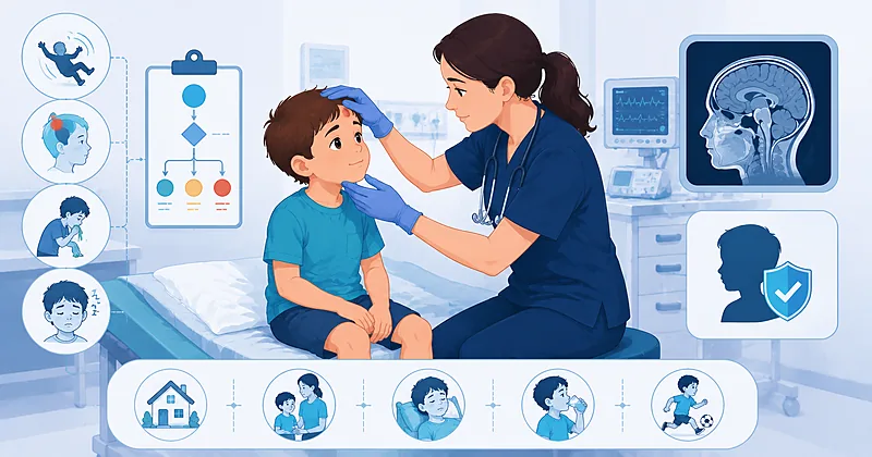
- 1Apply the PECARN rule only to children with a Glasgow Coma Scale of 14–15 and no penetrating trauma — it was never designed for severe injury.
- 2Use a different rule set for each age band — the under-2 and 2-and-older pathways have distinct predictors.
- 3A child with none of the PECARN high-risk predictors has a risk of clinically important brain injury well under 0.05% — CT can be safely avoided.
- 4Reserve immediate CT for the highest-risk children: altered mental status or palpable skull fracture (under 2), or signs of basilar skull fracture (2 and older).
- 5For the intermediate-risk child, choose between CT and structured observation rather than scanning reflexively.
- 6A period of observation of 4–6 hours often resolves intermediate-risk cases without imaging as symptoms declare themselves.
- 7Never order skull radiographs to rule out intracranial injury — they neither confirm nor exclude clinically important findings.
- 8Discharge with clear written return precautions; most concussion symptoms resolve within one to four weeks with reassurance and graded return to activity.
Who Counts as a Pediatric Minor Head Injury
Before any decision rule applies, the child must actually fit the definition. A pediatric minor head injury means blunt trauma in a child who arrives alert and responsive, with a Glasgow Coma Scale of 14 or 15 and no focal neurological deficit. Children outside this window — those with penetrating injury, a GCS of 13 or below, or known structural brain conditions — fall outside the validated pathway and warrant individualized, often more aggressive, evaluation.
Evaluate every child for inclusion before applying a decision rule: confirm blunt mechanism, a GCS of 14–15, and the absence of focal deficits. The PECARN rule loses its safety guarantees outside these boundaries.
Strong Rec High Evidence PECARN 2009Evaluate for non-accidental trauma in any infant with an inconsistent history, injury inconsistent with developmental stage, or unexplained findings — this overrides routine low-risk pathways.
Moderate Rec Low Evidence CDC 2018Pediatric Minor Head Injury Imaging in Children Under 2
The youngest children are the hardest to assess and the most radiosensitive, which is exactly why the PECARN rule built a separate pathway for them. In a pediatric minor head injury in a child under 2, the highest-risk features are altered mental status and a palpable skull fracture. Their presence pushes toward immediate imaging; their absence opens the door to a far more measured approach.
Perform CT promptly in a child under 2 with altered mental status or a palpable skull fracture. These two findings carry the highest probability of clinically important traumatic brain injury in this age band.
Strong Rec High Evidence PECARN 2009Consider observation versus CT in a child under 2 with intermediate-risk features: an occipital, parietal, or temporal scalp hematoma, loss of consciousness of five seconds or more, a severe mechanism, or behavior judged abnormal by the parent.
Conditional Rec Moderate Evidence PECARN 2009Do not routinely image a child under 2 who is acting normally, has a normal mental status, no scalp hematoma except a small frontal one, and none of the other intermediate-risk features. The risk of important injury approaches background levels.
Strong Rec High Evidence PECARN 2009Imaging Decisions in Children 2 and Older
Older children can describe symptoms and cooperate with examination, which shifts the predictor set. Here the two highest-risk findings are altered mental status and clinical signs of a basilar skull fracture. The intermediate tier — loss of consciousness, vomiting, severe mechanism, or severe headache — again becomes the zone where observation earns its place.
Perform CT promptly in a child 2 or older with altered mental status or signs of a basilar skull fracture such as hemotympanum, periorbital or postauricular bruising, or cerebrospinal fluid leak.
Strong Rec High Evidence PECARN 2009Consider CT versus a period of observation in a child 2 or older with any single intermediate-risk feature: a history of loss of consciousness, vomiting, a severe injury mechanism, or severe headache.
Conditional Rec Moderate Evidence PECARN 2009Do not image a child 2 or older with a normal mental status and none of the high- or intermediate-risk predictors. Reassurance and discharge with return precautions is the evidence-supported path.
Strong Rec High Evidence PECARN 2009Do not order skull radiographs to screen for intracranial injury in any age band. A normal film does not exclude brain injury, and an abnormal one does not change acute management.
Against Moderate Evidence CDC 2018Clinical Decision Pathway
A practical, question-based approach to the child in front of you. Work through the questions in order rather than jumping to a scan.
Predictor Reference by Age and Risk Tier
This reference is organized by age band and risk tier with an added column for the practical action each tier triggers — a structure built for bedside use rather than research reporting.
| Age Band | High-Risk (CT Now) | Intermediate (CT or Observe) | Practical Action |
|---|---|---|---|
| Under 2 years | Altered mental status; palpable skull fracture | Non-frontal scalp hematoma; LOC ≥5 s; severe mechanism; not acting normally per parent | Engage caregiver early; observation is often preferable to sedation for CT |
| 2 years and older | Altered mental status; basilar skull fracture signs | Any LOC; vomiting; severe mechanism; severe headache | Count the features — a single isolated finding favors observation |
| Both bands | GCS <14 or focal deficit (outside rule) | Anticoagulation, bleeding disorder, or shunt — lower threshold | Treat these as out-of-rule; individualize the decision |
- Severe mechanism includes high-speed motor vehicle events, falls of significant height for age, or being struck by a high-impact object.
- The “acting normally” judgment relies heavily on the caregiver who knows the child — take it seriously.
Monitoring and Follow-Up
Whether a child is observed or discharged, the follow-up plan matters as much as the initial decision. This schedule is framed around what to watch, when, and the pitfall that most often trips clinicians up.
| What to Monitor | When | What to Look For | Common Pitfall |
|---|---|---|---|
| ED observation | 4–6 hours from injury | Improving alertness, tolerating fluids, no new vomiting | Discharging at hour 2 before symptoms have had time to declare |
| Return precautions | At discharge, in writing | Repeated vomiting, worsening headache, confusion, seizure, unequal pupils | Verbal-only instructions that caregivers forget under stress |
| Concussion recovery | Days to weeks | Symptom resolution; graded return to learn and play | Prolonged strict rest, which can delay rather than speed recovery |
Evidence in Context
What the underlying studies actually showed, where the major bodies align, and where judgment still carries the decision.
How the prediction rules were derived and validated
The original work enrolled tens of thousands of children with blunt head trauma across many emergency departments, then derived and prospectively validated age-specific predictors for clinically important brain injury. The headline result is the high sensitivity of the low-risk criteria, which is what makes safely withholding a CT defensible.
Why radiation exposure drives the whole strategy
The developing brain is more vulnerable to ionizing radiation, and the lifetime cancer risk from a head CT is small but not zero, and is concentrated in the youngest patients. The entire decision framework exists to keep that cumulative exposure as low as is consistent with not missing a dangerous injury.
The value of observation in the intermediate group
Subsequent analyses found that a strategy of watching intermediate-risk children rather than scanning them immediately reduced CT use without increasing missed injuries. Symptoms that are going to evolve usually do so within a few hours, which is the rationale behind a structured observation window.
Where guidance converges on managing concussion
National guidance broadly agrees that most children with concussion recover within one to four weeks, that prolonged strict rest is unhelpful, and that imaging plays no role in routine concussion diagnosis. The convergence on graded return to activity is now strong across sources.
References
- 1.Kuppermann N, Holmes JF, Dayan PS, et al. Identification of children at very low risk of clinically-important brain injuries after head trauma: a prospective cohort study. Lancet. 2009;374(9696):1160–1170. doi:10.1016/S0140-6736(09)61558-0
- 2.Lumba-Brown A, Yeates KO, Sarmiento K, et al. Centers for Disease Control and Prevention Guideline on the Diagnosis and Management of Mild Traumatic Brain Injury Among Children. JAMA Pediatr. 2018;172(11):e182853. doi:10.1001/jamapediatrics.2018.2853
- 3.Nigrovic LE, Schunk JE, Foerster A, et al. The effect of observation on cranial computed tomography utilization for children after blunt head trauma. Pediatrics. 2011;127(6):1067–1073. doi:10.1542/peds.2010-3373
- 4.Babl FE, Borland ML, Phillips N, et al. Accuracy of clinical decision rules for children with minor head injury: a prospective cohort study (APHIRST). Lancet. 2017;389(10087):2393–2402. doi:10.1016/S0140-6736(17)30555-X
How to Read the Evidence Tags
Each recommendation carries a strength tag and an evidence-quality tag — Medaptly’s own simplified interpretations, not any single guideline body’s classification system.
Recommendation Strength
| Tag | What It Means |
|---|---|
| Strong Rec | High-quality evidence broadly supports this action. |
| Moderate Rec | The weight of evidence favours this action. |
| Conditional Rec | The benefit is less certain — individualise to the patient. |
| Against | Evidence shows no benefit or potential harm. |
Evidence Quality
| Tag | What It Means |
|---|---|
| High Evidence | Multiple well-designed studies or high-quality prospective cohorts. |
| Moderate Evidence | Single cohort or large observational studies. |
| Low Evidence | Expert consensus or smaller studies. |