
Abdominal Aortic Aneurysm: 8 Essential 2026 Rules
Clinical Practice Update — Screening, Surveillance, and Repair Strategy in Adults
This is an original clinical education article informed by current guidelines and evidence. See References below for source documents.
- Clinical Focus
- Evidence-based abdominal aortic aneurysm screening, surveillance, and repair in adults
- Target Audience
- Primary care physicians, vascular surgeons, emergency physicians, radiologists, residents
- Setting
- Primary care screening, vascular clinic surveillance, emergency department, operating theatre
- Source Evidence
- •USPSTF Recommendation on AAA Screening (2019)
- •Society for Vascular Surgery (SVS) Practice Guidelines on AAA (2018)
- •European Society for Vascular Surgery (ESVS) Guidelines on AAA (2019)
- •IMPROVE Trial — Endovascular vs Open Repair of Ruptured AAA (BMJ, 2017)
- •EVAR-1 Trial Long-Term Follow-Up (Lancet, 2016)
Key Clinical Takeaways
Effective abdominal aortic aneurysm care in 2026 still hinges on three decisions made at different points in the patient journey: whom to screen, when to repair, and how to follow up. The points below distil the evidence into actionable rules.
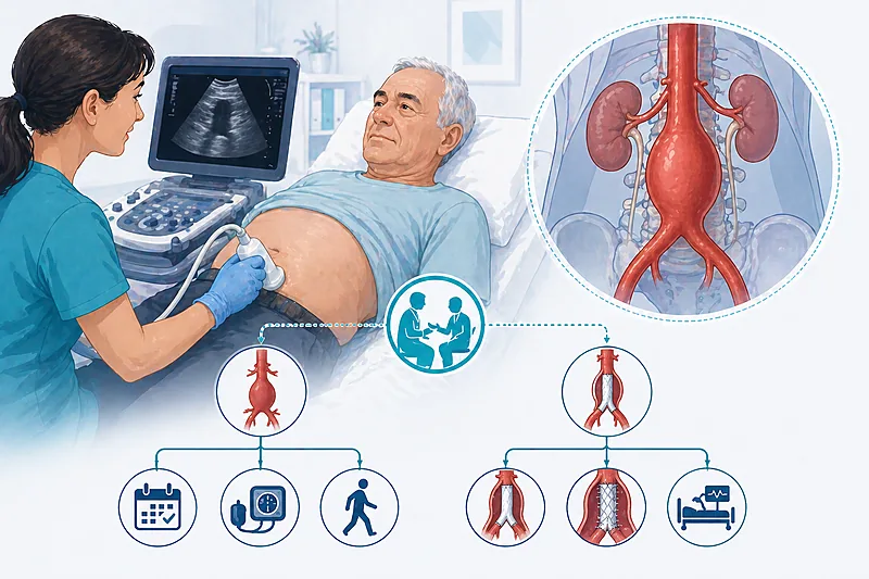
- 1Offer one-time abdominal ultrasound to every man aged 65–75 who has ever smoked → Screening
- 2Match surveillance frequency to sac diameter — a 3.5 cm sac does not need the same interval as a 5.0 cm sac → Surveillance
- 3Offer elective repair at 5.5 cm in men and 5.0 cm in women, or at rapid expansion ≥1 cm per year → Repair Indications
- 4Choose EVAR for anatomically suitable patients with higher operative risk — lower 30-day mortality → Repair Technique
- 5Consider open repair for younger, fitter patients with a long expected life span — better long-term durability → Repair Technique
- 6In ruptured AAA, EVAR first if anatomically suitable — no mortality disadvantage vs open → Ruptured AAA
- 7Book CT angiography at 30 days post-EVAR, then annual imaging indefinitely → Post-EVAR
- 8Counsel every patient on smoking cessation — the single most effective intervention for slowing expansion → Medical Therapy
Who Needs Screening for Abdominal Aortic Aneurysm?
One-time ultrasound is the cornerstone of abdominal aortic aneurysm screening because population-based trials show it reduces aneurysm-related mortality in older men with a smoking history. Both USPSTF and SVS frameworks endorse this, but they disagree slightly on whom to screen beyond the core group.
Perform a one-time screening abdominal ultrasound in all men aged 65–75 with any history of tobacco smoking, whether current or former.
Strong Rec High Evidence USPSTF 2019 SVS 2018Consider selective screening in men aged 65–75 who have never smoked but have a first-degree relative with abdominal aortic aneurysm, reflecting a familial risk that approaches one in five.
Conditional Rec Moderate Evidence SVS 2018Evaluate for screening in women aged 65–75 who have smoked or have a positive family history — the evidence base is thinner than for men, and shared decision-making is essential.
Conditional Rec Low Evidence USPSTF 2019Do not repeat routine screening in patients whose initial ultrasound showed an aortic diameter below 3.0 cm — the risk of clinically important abdominal aortic aneurysm developing later is very low.
Against Moderate Evidence SVS 2018Surveillance of Small Abdominal Aortic Aneurysm
Once a small abdominal aortic aneurysm is detected, the surveillance interval should scale with diameter. A sac measuring 3.0–3.9 cm expands slowly; a 5.0–5.4 cm sac can tip into repair territory within months.
Use ultrasound as the first-line surveillance modality in small and medium aneurysms — no radiation, low cost, and adequate reproducibility for serial diameter measurements.
Strong Rec High Evidence SVS 2018 ESVS 2019Refer to vascular surgery for assessment once the aortic diameter reaches 5.0 cm in men and 4.5 cm in women — this gives time to optimise comorbidities before the repair threshold is reached.
Strong Rec Moderate Evidence SVS 2018Counsel patients that rapid expansion — defined as ≥0.5 cm over six months or ≥1.0 cm over a year — prompts earlier referral regardless of absolute diameter.
Moderate Rec Moderate Evidence ESVS 2019Surveillance Intervals by Sac Diameter
| Sac Diameter | Ultrasound Interval | What to Flag to Patient | Common Pitfalls |
|---|---|---|---|
| 3.0–3.9 cm | Every 3 years | Low-risk — lifestyle first | Losing track of recall — set a diary |
| 4.0–4.4 cm | Every 12 months | Start BP and statin optimisation | Forgetting to address tobacco use |
| 4.5–4.9 cm | Every 6 months | Refer women to vascular surgery | Using male thresholds in women |
| 5.0–5.4 cm | Every 3–6 months | Refer all men — plan elective repair | Delaying referral hoping for stability |
| ≥5.5 cm (men) / ≥5.0 cm (women) | CT angiography | At repair threshold | Repeating ultrasound instead of CT |
Elective Repair: When to Fix an Abdominal Aortic Aneurysm
The decision to proceed with elective abdominal aortic aneurysm repair balances rupture risk against operative mortality. At 5.5 cm in men the annual rupture risk climbs sharply, and most patients cross into a zone where the risk of leaving the aneurysm in place exceeds the risk of fixing it.
Offer elective repair to men with an infrarenal abdominal aortic aneurysm of 5.5 cm or greater, provided perioperative risk is acceptable.
Strong Rec High Evidence SVS 2018 ESVS 2019Offer elective repair to women at a lower diameter threshold of 5.0 cm, because the absolute aortic size at which rupture occurs is smaller in female patients.
Moderate Rec Moderate Evidence SVS 2018 ESVS 2019Consider earlier repair below the size threshold when rapid expansion, symptoms attributable to the aneurysm, or a saccular morphology is documented.
Moderate Rec Moderate Evidence SVS 2018Do not offer routine elective repair in patients whose life expectancy is short (under two years) because of unrelated comorbidities — the survival benefit does not accrue.
Against Low Evidence SVS 2018Prescribe moderate- to high-intensity statin therapy and a single antiplatelet agent before elective repair — these medical measures reduce perioperative cardiovascular events.
Strong Rec High Evidence SVS 2018Choosing Between EVAR and Open Repair
Both techniques are effective; the decision rests on anatomy, age, operative risk, and the patient’s willingness to commit to lifelong imaging follow-up. EVAR offers lower early mortality, open repair offers greater durability.
Offer EVAR as the first-line technique for most anatomically suitable patients — 30-day mortality is roughly a third of that seen with open repair.
Strong Rec High Evidence SVS 2018 ESVS 2019Consider open repair in younger, fitter patients with a long expected survival — late reintervention rates are lower and lifelong imaging is not required.
Moderate Rec High Evidence EVAR-1 2016 SVS 2018Counsel patients opting for EVAR that they will require lifelong surveillance imaging and have a roughly 20–30% chance of reintervention over a decade.
Strong Rec High Evidence EVAR-1 2016Do not offer EVAR when the anatomy is hostile — short or angulated proximal necks, heavily calcified iliac vessels, or small access arteries all predict early endoleak and device failure.
Against Moderate Evidence SVS 2018EVAR vs Open Repair: A Patient-Profile Comparison
| Patient Profile | Preferred Technique | Rationale | Watch-Out |
|---|---|---|---|
| Elderly, high operative risk, favourable anatomy | EVAR | Low 30-day mortality, fast recovery | Compliance with lifelong imaging |
| Younger (<70), fit, long life expectancy | Open | Lower reintervention, no lifelong imaging | Higher short-term morbidity |
| Hostile proximal neck or iliac anatomy | Open (or fenestrated EVAR) | Standard EVAR likely to fail | Need centre experienced in complex cases |
| Juxta- or para-renal aneurysm | Fenestrated/branched EVAR or open | Requires visceral vessel preservation | Centralise to tertiary unit |
| Connective tissue disorder (e.g. Marfan) | Open | Friable tissue — endograft seal uncertain | Genetic evaluation of relatives |
Ruptured Abdominal Aortic Aneurysm: Emergency Management
Ruptured abdominal aortic aneurysm is a time-critical emergency. The classic triad of back or flank pain, hypotension, and a pulsatile abdominal mass is incomplete in many patients — a low threshold for imaging in older patients with unexplained collapse is lifesaving.
Start immediate CT angiography for suspected rupture in a haemodynamically stable patient — confirms the diagnosis and maps anatomy for possible EVAR.
Strong Rec High Evidence IMPROVE 2017 ESVS 2019Perform permissive hypotension (target systolic BP 70–90 mmHg, preserving consciousness) before definitive repair — aggressive fluid resuscitation worsens haemorrhage.
Strong Rec Moderate Evidence ESVS 2019Offer EVAR as the first-line emergency repair technique for ruptured abdominal aortic aneurysm when anatomy permits — IMPROVE and long-term follow-up show non-inferior mortality and lower early morbidity than open surgery.
Strong Rec High Evidence IMPROVE 2017Transfer haemodynamically unstable patients directly to theatre — waiting for imaging in a collapsed patient with a known AAA loses more than it gains.
Moderate Rec Low Evidence ESVS 2019Post-EVAR Surveillance and Endoleak Management
Every patient who undergoes EVAR for an abdominal aortic aneurysm enters a lifelong surveillance relationship. Device migration, sac expansion, and endoleak can all occur silently years after an apparently successful repair.
Arrange CT angiography at 30 days following EVAR, with subsequent imaging annually — delayed sac expansion is the most common indicator of occult endoleak.
Strong Rec Moderate Evidence SVS 2018Treat Type I and Type III endoleak promptly — both represent direct systemic pressurisation of the sac and carry a substantial rupture risk.
Strong Rec High Evidence SVS 2018 ESVS 2019Avoid routine intervention for Type II endoleak alone — most are self-limiting; intervene only when associated with sac expansion >5 mm.
Conditional Rec Moderate Evidence SVS 2018Consider duplex ultrasound as an alternative to CT in selected stable patients after year two, reducing cumulative radiation and contrast exposure.
Conditional Rec Moderate Evidence ESVS 2019Endoleak Types at a Glance
| Type | Source of Flow | Clinical Action | Urgency |
|---|---|---|---|
| I | Inadequate seal at proximal (Ia) or distal (Ib) attachment | Repair — proximal cuff, extension, or conversion | High |
| II | Retrograde flow from lumbar or inferior mesenteric vessels | Observe; embolise only if sac expands | Low |
| III | Structural failure between graft components or fabric tear | Repair urgently — relining or conversion | High |
| IV | Fabric porosity (rare with modern grafts) | Usually resolves — observe | Low |
| V (endotension) | Sac pressurisation without demonstrable leak | Consider relining if expansion occurs | Moderate |
Medical Management During Surveillance
Patients with a small aneurysm under surveillance are almost always at high cardiovascular risk. The medical therapy plan is not primarily about slowing aneurysm expansion — it is about keeping the patient alive and well enough to benefit from eventual repair.
Optimise blood pressure control to a target below 130/80 mmHg in every patient with an abdominal aortic aneurysm.
Strong Rec Moderate Evidence SVS 2018Prescribe a statin in every patient with an abdominal aortic aneurysm, regardless of baseline LDL, for cardiovascular event reduction.
Strong Rec High Evidence SVS 2018Counsel every current smoker on cessation at each visit — continued smoking roughly doubles the rate of sac expansion and trebles rupture risk.
Strong Rec High Evidence USPSTF 2019 SVS 2018Do not prescribe antibiotics or doxycycline to slow aneurysm expansion — trials have failed to demonstrate a benefit.
Against High Evidence SVS 2018Clinical Decision Pathway
A practical, question-based approach to managing a patient with a known or suspected abdominal aortic aneurysm. Work through the questions in order.
Monitoring and Follow-Up
| Parameter | When to Check | Action Threshold | Common Pitfalls |
|---|---|---|---|
| Sac diameter (ultrasound) | Interval scaled to size | ≥1 cm/year growth or crossing repair threshold | Using different operators without cross-calibration |
| Blood pressure | Every clinic visit | Target <130/80 mmHg | Failing to titrate in elderly — start low, go slow but do titrate |
| Tobacco status | Every visit | Any continued use → intervention | “They know they should stop” — brief advice still works |
| Post-EVAR CT | 30 days, then annually | Sac growth ≥5 mm or new endoleak | Losing patients to follow-up between cardiology, GP, and vascular teams |
| Lipid profile | At diagnosis and 12-monthly | LDL-C <1.8 mmol/L (on statin) | Stopping statin because of non-specific aches |
Evidence in Context
What the trials show, where the major guidelines agree, and where they diverge.
Where SVS and ESVS Agree
Both frameworks agree on: ultrasound as the first-line screening and surveillance modality, the 5.5 cm diameter threshold for elective repair in men, EVAR as the preferred technique for most anatomically suitable patients, and the need for lifelong post-EVAR imaging.
Where SVS and ESVS Differ
Screening of women: ESVS takes a more permissive view, supporting screening in selected women with risk factors; SVS remains more cautious because the evidence base in women is thinner.
Both documents acknowledge this gap honestly — it reflects limited trial data, not a disagreement about biology.
What the IMPROVE Trial Showed
IMPROVE compared endovascular with open strategies for ruptured AAA in anatomically suitable patients. At 30 days, mortality was similar; at 3 years, the endovascular strategy was associated with better survival and cost-effectiveness. The take-home is that EVAR-first is a reasonable strategy when anatomy and logistics allow.
Long-Term Lessons from EVAR-1
The long-term follow-up of EVAR-1 demonstrated that the early mortality advantage of EVAR over open repair diminishes over time, with late graft-related complications eroding the benefit. This underpins the current preference for open repair in younger, fitter patients.
Evidence Gaps We Still Face
The optimal diameter threshold for elective repair in women remains undefined — most evidence extrapolates from male-dominated trials. Trials of medical therapy for aneurysm expansion have been consistently disappointing, and there is no drug with a proven role in shrinking or stabilising a known aneurysm.
References
- 1. US Preventive Services Task Force. Screening for Abdominal Aortic Aneurysm: US Preventive Services Task Force Recommendation Statement. JAMA. 2019;322(22):2211–2218. doi:10.1001/jama.2019.18928
- 2. Chaikof EL, Dalman RL, Eskandari MK, et al. The Society for Vascular Surgery practice guidelines on the care of patients with an abdominal aortic aneurysm. J Vasc Surg. 2018;67(1):2–77.e2. doi:10.1016/j.jvs.2017.10.044
- 3. Wanhainen A, Verzini F, Van Herzeele I, et al. Editor’s Choice — European Society for Vascular Surgery (ESVS) 2019 Clinical Practice Guidelines on the Management of Abdominal Aorto-iliac Artery Aneurysms. Eur J Vasc Endovasc Surg. 2019;57(1):8–93. doi:10.1016/j.ejvs.2018.09.020
- 4. IMPROVE Trial Investigators. Comparative clinical effectiveness and cost effectiveness of endovascular strategy v open repair for ruptured abdominal aortic aneurysm: three year results of the IMPROVE randomised trial. BMJ. 2017;359:j4859. doi:10.1136/bmj.j4859
- 5. Patel R, Sweeting MJ, Powell JT, Greenhalgh RM; EVAR trial investigators. Endovascular versus open repair of abdominal aortic aneurysm in 15-years’ follow-up of the UK endovascular aneurysm repair trial 1 (EVAR trial 1). Lancet. 2016;388(10058):2366–2374. doi:10.1016/S0140-6736(16)31135-7
How to Read the Evidence Tags
Every recommendation in this article carries two tags — recommendation strength and evidence quality. These are Medaptly’s simplified interpretations, designed for bedside use.
Recommendation Strength
| Tag | What It Means |
|---|---|
| Strong Rec | High-quality evidence broadly supports this action. |
| Moderate Rec | Evidence favours this action for most patients. |
| Conditional Rec | Benefit is less certain — individualise based on patient factors. |
| Against | Evidence shows no benefit or potential harm. |
Evidence Quality
| Tag | What It Means |
|---|---|
| High Evidence | Multiple well-designed RCTs or high-quality meta-analyses. |
| Moderate Evidence | Single RCT or large observational studies. |
| Low Evidence | Expert consensus or small studies. |