
Hyperparathyroidism Surgery: 7 Proven Localization Rules
Clinical Practice Update — Localization, Intraoperative PTH Monitoring, and Reoperation Strategy
This is an original clinical education article informed by current guidelines and evidence. See References below for source documents.
- Clinical Focus
- Evidence-based hyperparathyroidism surgery covering localization studies, IOPTH monitoring, and reoperation
- Target Audience
- General surgeons, endocrine surgeons, otolaryngologists, endocrinologists, surgical trainees
- Setting
- Endocrine surgery clinics, ambulatory surgical centers, tertiary referral centers
- Source Evidence
- •AAES Guidelines for Definitive Management of Primary Hyperparathyroidism (2016)
- •Fifth International Workshop on Primary Hyperparathyroidism (2022)
- •European Society of Endocrine Surgeons Consensus on Reoperative Parathyroidectomy
- •Irvin GL et al. Miami Criterion for IOPTH (Am J Surg)
Key Clinical Takeaways
Effective hyperparathyroidism surgery hinges on three sequential decisions: confirming the biochemical diagnosis, localizing the abnormal gland or glands, and verifying cure intraoperatively. The points below distill these three pillars into bedside-ready actions.
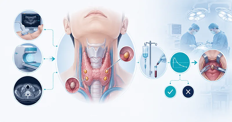
- 1Confirm the biochemical diagnosis before ordering any imaging — hyperparathyroidism is diagnosed in the laboratory, not on the scan
- 2Pair two complementary modalities — high-resolution neck ultrasound plus sestamibi with SPECT/CT — for first-time hyperparathyroidism surgery
- 3Reach for 4D-CT when first-line imaging is negative or discordant — not as the routine starting point
- 4Apply the Miami criterion — a greater-than-50% PTH drop at 10 minutes post-excision from the highest pre-excision baseline — to confirm intraoperative cure
- 5Convert to bilateral neck exploration when intraoperative PTH does not drop adequately — multi-gland disease is the usual culprit
- 6For reoperation, reconfirm biochemistry, review the prior operative note and pathology, and obtain two concordant imaging modalities
- 7Refer every reoperative case to a high-volume parathyroid surgeon — complication rates rise sharply outside specialist hands
- 8Reserve selective venous sampling for failed reoperative imaging at experienced centers — it is a salvage tool, not a screen
- 9Counsel reoperative patients on substantially higher risks of permanent hypoparathyroidism and recurrent laryngeal nerve injury
- 10Confirm biochemical cure with calcium and PTH at six months — not before — and monitor annually thereafter
Selecting Patients for Hyperparathyroidism Surgery
Hyperparathyroidism surgery is curative in over 95% of first-time cases when performed by experienced surgeons. The first task is identifying who needs it. Symptomatic patients are easy — the harder conversation is around asymptomatic disease, where surgical criteria have been refined over five international workshops.
Refer all adults with symptomatic primary hyperparathyroidism for surgical evaluation, including those with nephrolithiasis, fragility fractures, neuromuscular symptoms, or hypercalcemic crisis.
Strong Rec High Evidence AAES 2016Refer asymptomatic adults meeting any single workshop criterion: serum calcium more than 1 mg/dL above the upper reference limit, eGFR below 60 mL/min, lumbar spine, total hip or distal radius T-score at or below −2.5, vertebral fracture on imaging, 24-hour urinary calcium above 400 mg with elevated stone risk, or age younger than 50 years.
Strong Rec High Evidence IWPHPT 2022Consider hyperparathyroidism surgery in normocalcemic primary hyperparathyroidism when end-organ effects (stones, low bone density, neurocognitive symptoms) are documented and secondary causes have been excluded.
Conditional Rec Moderate Evidence IWPHPT 2022Discontinue thiazide diuretics and lithium for 4–8 weeks (when clinically feasible) and recheck calcium and PTH before confirming the diagnosis.
Strong Rec Moderate Evidence AAES 2016Measure 25-hydroxyvitamin D in every candidate for hyperparathyroidism surgery and replete to a level above 30 ng/mL preoperatively to reduce the risk of postoperative hungry bone syndrome.
Moderate Rec Moderate Evidence AAES 2016Screen for familial syndromes (MEN1, MEN2A, MEN4, familial isolated hyperparathyroidism, hyperparathyroidism-jaw tumor syndrome) when patients are younger than 40, have multi-gland disease, or have a suggestive family history.
Strong Rec Moderate Evidence AAES 2016Localization Studies Before Hyperparathyroidism Surgery
Imaging in hyperparathyroidism surgery answers one question only: where is the abnormal gland? It does not answer whether the patient has the disease. Once the surgical decision is made, the role of localization is to enable a focused operation while preserving the option to convert to bilateral exploration.
Do not order localization imaging until the biochemical diagnosis is confirmed and the patient is a surgical candidate — imaging has no diagnostic role in primary hyperparathyroidism.
Strong Rec High Evidence AAES 2016Obtain high-resolution neck ultrasound as the initial localization study before first-time hyperparathyroidism surgery. Ultrasound also evaluates concurrent thyroid pathology, which is found in roughly one in three patients.
Strong Rec High Evidence AAES 2016Perform a sestamibi scan with SPECT/CT as a complementary modality — concordance between ultrasound and sestamibi predicts single-adenoma disease with very high accuracy and supports a focused approach.
Strong Rec Moderate Evidence AAES 2016Order 4D-CT when ultrasound and sestamibi are negative, discordant, or when an ectopic gland (mediastinal, retro-esophageal, intrathyroidal) is suspected. 4D-CT offers the highest sensitivity for small or ectopic adenomas.
Moderate Rec Moderate Evidence AAES 2016Consider 18F-fluorocholine PET/CT when conventional imaging fails before considering reoperation — emerging evidence suggests it outperforms sestamibi in localizing small or hyperplastic glands.
Conditional Rec Moderate Evidence ESES ConsensusDo not perform fine-needle aspiration of suspected parathyroid lesions — it does not change management, can cause local fibrosis that complicates surgery, and risks seeding.
Against Moderate Evidence AAES 2016Choosing a Localization Modality — A Practical Guide
The table below organizes the available modalities by clinical scenario rather than by modality alone, so the right test is the easy choice at the point of decision-making.
| Clinical Scenario | First-Line Imaging | If Negative or Discordant | Practical Tip |
|---|---|---|---|
| First-time surgery, classic biochemistry | Ultrasound + sestamibi SPECT/CT | 4D-CT | Concordant pair = focused approach |
| Suspected multi-gland disease (mild biochemistry, lithium use) | Plan for bilateral exploration | Imaging less critical | IOPTH guides extent of resection |
| Coexisting thyroid nodules | Ultrasound | Add sestamibi SPECT/CT | Address thyroid pathology concurrently |
| Suspected ectopic gland | 4D-CT (chest + neck) | 18F-fluorocholine PET/CT | Image mediastinum thoroughly |
| Reoperation | 4D-CT + sestamibi SPECT/CT (two concordant) | 18F-fluorocholine PET/CT, then selective venous sampling | Never reoperate on a single positive scan |
| Pregnancy or iodine contrast allergy | Ultrasound | MRI (non-contrast) | Defer non-essential imaging in pregnancy |
Intraoperative PTH Monitoring During Hyperparathyroidism Surgery
Intraoperative parathyroid hormone (IOPTH) monitoring is the in-theatre confirmation that hyperparathyroidism surgery has achieved its goal. PTH has a half-life of roughly 3–5 minutes, so a meaningful drop after excising an abnormal gland is detectable within 10 minutes. Multiple criteria exist, but the Miami criterion has become the workhorse.
Apply the Miami criterion as the default interpretation rule: a drop of more than 50% from the highest pre-excision baseline, measured at 10 minutes after gland removal. This predicts long-term cure in approximately 97–98% of cases.
Strong Rec High Evidence Irvin et al. Am J SurgDraw a baseline PTH sample at induction and a second baseline immediately before suspect-gland excision. Use the higher of the two as the comparator — gland manipulation often raises PTH transiently.
Strong Rec High Evidence AAES 2016Continue neck exploration if the 10-minute post-excision PTH does not meet the Miami threshold — the residual hypersecretion almost always comes from another abnormal gland.
Strong Rec High Evidence AAES 2016Send an additional sample at 15 or 20 minutes when the 10-minute drop is borderline (between 40% and 50%) — some patients show a delayed but adequate decline.
Moderate Rec Moderate Evidence AAES 2016Aim for the final intraoperative PTH to fall within the normal reference range whenever feasible — an in-range final value adds a layer of biochemical confidence beyond percent drop alone.
Moderate Rec Moderate Evidence ESES ConsensusRecognize the false-positive scenario: a single adenoma can mask a second abnormal gland, and the 10-minute drop may appear adequate while a second adenoma continues to secrete. If preoperative biochemistry was very mild, lower the threshold for additional exploration.
Moderate Rec Low Evidence ESES ConsensusDo not interpret a rapid early drop as a license to terminate exploration without the 10-minute confirmation. Premature closure is one of the most common avoidable causes of operative failure.
Against Moderate Evidence Carneiro et al.Use IOPTH routinely in bilateral exploration as well as focused approaches — it identifies double adenomas missed by visual inspection alone.
Strong Rec Moderate Evidence AAES 2016IOPTH Criteria at a Glance
| Criterion | Drop Threshold | Baseline Reference | When to Use | Trade-Off |
|---|---|---|---|---|
| Miami | >50% at 10 min | Highest pre-excision value | Default in most centers | May miss subtle multi-gland disease |
| Halle | Drop to normal range | Lab normal reference | Stricter biochemical cure | More re-explorations; higher false-negative rate |
| Rome | >50% & in normal range at 20 min | Pre-incision value | Multi-gland-prone cases | Longer operative time |
| Vienna | >50% at 10 min | Pre-incision value | Centers using single baseline | Misses manipulation-related spikes |
Reoperation Strategy After Failed Surgery
Persistent hyperparathyroidism (failure to achieve normocalcemia within 6 months) and recurrent hyperparathyroidism (relapse after at least 6 months of normocalcemia) require a fundamentally different approach. Cure rates fall to roughly 80–95% in experienced centers, and complication rates rise. The single most important decision is who should perform the operation.
Refer every reoperative candidate to a high-volume parathyroid surgeon (typically defined as more than 50 parathyroid cases annually) at a tertiary center.
Strong Rec High Evidence AAES 2016Reconfirm the biochemical diagnosis before any reoperation. Exclude familial hypocalciuric hypercalcemia with a calcium-to-creatinine clearance ratio and consider genetic testing where appropriate.
Strong Rec High Evidence AAES 2016Obtain the prior operative report, surgical pathology, and original imaging before planning reoperation. The location of glands previously identified, removed, or biopsied determines the most efficient surgical approach.
Strong Rec High Evidence AAES 2016Require two concordant imaging modalities before re-exploring the neck. 4D-CT plus sestamibi SPECT/CT is the most commonly used combination at experienced centers.
Strong Rec Moderate Evidence ESES ConsensusConsider 18F-fluorocholine PET/CT as the next step when conventional imaging is non-localizing or discordant before resorting to invasive studies.
Moderate Rec Moderate Evidence ESES ConsensusReserve selective venous sampling for reoperative cases with non-localizing non-invasive imaging at centers experienced in the technique. It is a salvage tool, not a routine investigation.
Conditional Rec Moderate Evidence ESES ConsensusCounsel patients explicitly on the elevated risks of reoperation: permanent hypoparathyroidism around 3–5%, recurrent laryngeal nerve injury around 2–6%, and operative failure of 10–20% even in experienced hands.
Strong Rec High Evidence AAES 2016Perform preoperative laryngoscopy to document vocal cord function before reoperative hyperparathyroidism surgery — baseline assessment is essential for medico-legal and clinical follow-up.
Strong Rec Moderate Evidence AAES 2016Consider continuous intraoperative nerve monitoring in reoperative cases to reduce permanent recurrent laryngeal nerve injury, especially when re-entering a scarred operative field.
Moderate Rec Moderate Evidence ESES ConsensusPlan a targeted approach guided by imaging rather than routine bilateral re-exploration — the scar burden of a redo bilateral neck dissection drives complications without improving cure rates.
Strong Rec Moderate Evidence ESES ConsensusClinical Decision Pathway
A practical, question-based pathway through the evaluation, surgical planning, and intraoperative decision-making of a patient with confirmed primary hyperparathyroidism.
Monitoring and Follow-Up
Post-operative follow-up serves two purposes: catching the early complications of hypocalcemia and confirming durable biochemical cure. The schedule below condenses what to check, when, and what triggers action.
| Time Point | What to Check | Action Threshold | Common Pitfall |
|---|---|---|---|
| 6–24 hours postop | Serum calcium, symptoms of hypocalcemia | Symptomatic or calcium below 8.0 mg/dL → replete | Discharging before calcium has equilibrated |
| 1–2 weeks | Calcium, vitamin D, magnesium | Persistent hypocalcemia → suspect hungry bone syndrome | Underdosing calcium and active vitamin D in severe disease |
| 6 months | Calcium and PTH | Both normal → biochemical cure confirmed | Declaring cure too early — calcium can normalize before PTH |
| 12 months | DXA, 24-hour urinary calcium | BMD improvement expected at hip and spine | Failing to document the bone benefit of cure |
| Annually thereafter | Calcium and PTH | Rising values → consider recurrence | Discharge to PCP without a clear monitoring plan |
Start oral calcium 1–2 g daily for the first 2 weeks after hyperparathyroidism surgery in patients with significant preoperative disease (calcium above 12 mg/dL, severe osteoporosis, or markedly elevated PTH).
Moderate Rec Moderate Evidence AAES 2016Add calcitriol 0.25–0.5 mcg daily for patients at high risk of hungry bone syndrome — severe osteitis fibrosa, alkaline phosphatase markedly elevated, large adenoma.
Moderate Rec Moderate Evidence AAES 2016Confirm biochemical cure at 6 months — not earlier — with paired calcium and PTH measurements. Cure is defined as sustained normocalcemia beyond this point.
Strong Rec High Evidence AAES 2016Reassess bone mineral density 12 months after curative hyperparathyroidism surgery — significant gains at lumbar spine and hip are the expected outcome.
Moderate Rec Moderate Evidence IWPHPT 2022Monitor calcium and PTH annually for life after curative hyperparathyroidism surgery — recurrence remains possible decades later, particularly in patients with familial syndromes.
Moderate Rec Low Evidence AAES 2016Evidence in Context
Where the AAES, the International Workshop, and the European Society of Endocrine Surgeons agree, and where they part ways — with the practical implications for your operating list.
Where the major guidelines agree
All three bodies converge on the central pillars of hyperparathyroidism surgery: biochemical diagnosis comes before imaging, focused parathyroidectomy is appropriate when imaging is concordant, IOPTH monitoring improves cure rates, and reoperative cases belong at high-volume centers. The 50% threshold for the Miami criterion is the most widely adopted intraoperative rule across all three frameworks.
Where they differ — surgical criteria in asymptomatic disease
The AAES suggests considering surgery for any asymptomatic patient who is fit, even outside the formal workshop criteria, citing long-term bone and cardiovascular benefits. The International Workshop is more restrictive, surgery being firmly recommended only when at least one criterion is met. The practical implication: a 65-year-old fit patient with mild hypercalcemia and no end-organ effects could be reasonably referred under AAES guidance but observed under IWPHPT guidance.
Where they differ — the role of 18F-fluorocholine PET/CT
European centers have moved more decisively toward fluorocholine PET/CT as a second-line modality, with several prospective series showing superior sensitivity to sestamibi for small or hyperplastic glands. North American centers still lean on 4D-CT as the default second-line study, in part due to availability. The choice should be driven by which modality is well-performed locally, not by guideline geography.
IOPTH in negative imaging — what the trials show
Multiple series have demonstrated that IOPTH-guided bilateral exploration achieves cure rates comparable to focused parathyroidectomy when imaging fails to localize. The take-home is that negative imaging is not a contraindication to hyperparathyroidism surgery — it simply dictates a different operative strategy.
Near-infrared autofluorescence — emerging adjunct
Parathyroid tissue autofluoresces in the near-infrared spectrum, offering a label-free intraoperative method to identify glands. Early studies suggest reduced rates of inadvertent parathyroid resection during thyroid surgery and faster identification during reoperation. The technology is not yet a standard of care but is increasingly available in tertiary centers.
References
- 1.Wilhelm SM, Wang TS, Ruan DT, et al. The American Association of Endocrine Surgeons Guidelines for Definitive Management of Primary Hyperparathyroidism. JAMA Surg. 2016;151(10):959–968. doi:10.1001/jamasurg.2016.2310
- 2.Bilezikian JP, Khan AA, Silverberg SJ, et al. Evaluation and Management of Primary Hyperparathyroidism: Summary Statement and Guidelines from the Fifth International Workshop. J Bone Miner Res. 2022;37(11):2293–2314. doi:10.1002/jbmr.4677
- 3.Irvin GL 3rd, Dembrow VD, Prudhomme DL. Operative monitoring of parathyroid gland hyperfunction. Am J Surg. 1991;162(4):299–302. doi:10.1016/0002-9610(91)90135-Z
- 4.Carneiro DM, Solorzano CC, Nader MC, Ramirez M, Irvin GL 3rd. Comparison of intraoperative iPTH assays during parathyroidectomy for sporadic primary hyperparathyroidism. Surgery. 2003;134(6):973–979. doi:10.1016/j.surg.2003.06.001
- 5.Udelsman R, Lin Z, Donovan P. The superiority of minimally invasive parathyroidectomy based on 1650 consecutive patients with primary hyperparathyroidism. Ann Surg. 2011;253(3):585–591. doi:10.1097/SLA.0b013e318208fed9
- 6.Bergenfelz AOJ, Hellman P, Harrison B, Sitges-Serra A, Dralle H. Positional statement of the European Society of Endocrine Surgeons (ESES) on modern techniques in pHPT surgery. Langenbecks Arch Surg. 2009;394(5):761–764. doi:10.1007/s00423-009-0533-5
How to Read the Evidence Tags
Each recommendation carries two tags — one for recommendation strength, one for evidence quality — plus the source. These are Medaptly’s own simplified interpretations of the underlying source-grading systems.
Recommendation Strength
| Tag | What It Means |
|---|---|
| Strong Rec | High-quality evidence broadly supports this action. |
| Moderate Rec | The weight of evidence favours this action. |
| Conditional Rec | The benefit is less certain — individualize. |
| Against | Evidence shows no benefit or potential harm. |
Evidence Quality
| Tag | What It Means |
|---|---|
| High Evidence | Multiple well-designed RCTs or high-quality meta-analyses. |
| Moderate Evidence | Single RCT or large observational studies. |
| Low Evidence | Expert consensus or small studies. |