
Perforated Peptic Ulcer: From Resuscitation to Definitive Repair
Clinical Practice Update — Emergency Resuscitation, Risk Stratification, and Operative Repair of Perforated Peptic Ulcer in Adults
This is an original clinical education article informed by current guidelines and evidence. See References below for source documents.
- Clinical Focus
- Resuscitation, risk stratification, and operative perforated peptic ulcer repair in adults
- Target Audience
- General and acute care surgeons, surgical residents, emergency physicians, intensivists
- Setting
- Emergency department, operating room, surgical critical care
- Source Evidence
- •WSES Guidelines on Perforated and Bleeding Peptic Ulcer (World J Emerg Surg, 2020)
- •Surviving Sepsis Campaign — International Guidelines (Intensive Care Med, 2021)
- •Chan et al. Laparoscopic vs Open Omental Patch Repair Meta-Analysis (J Trauma Acute Care Surg, 2023)
- •PULP and Boey Risk Score Validation Studies (World J Surg; PMC, 2009–2020)
Key Clinical Takeaways
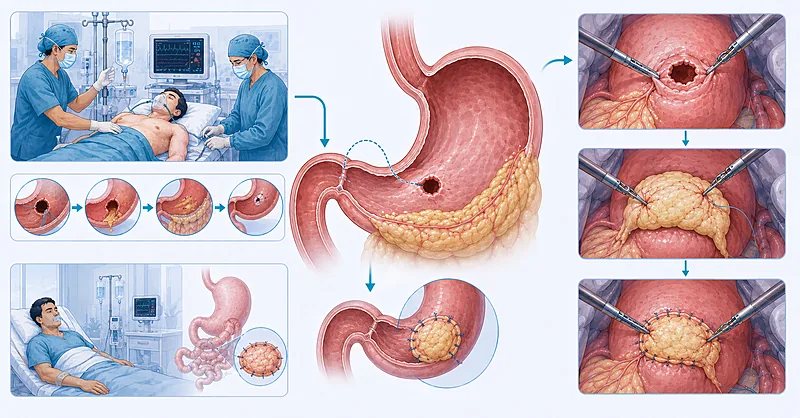
Successful perforated peptic ulcer repair begins long before the first incision. Outcomes hinge on three parallel priorities: aggressive physiologic resuscitation, rapid risk stratification, and prompt source control. A perforated peptic ulcer is a surgical emergency in which delay measurably raises mortality, so the resuscitation clock and the operative clock run together rather than in sequence. The points below distill the evidence into actionable rules for the bedside and the operating room.
- 1Start balanced crystalloid resuscitation and broad-spectrum antibiotics the moment perforation is suspected — do not wait for imaging confirmation → Resuscitation
- 2Screen for septic shock with qSOFA and lactate, and escalate to the operating room without delay in the deteriorating patient → Resuscitation
- 3Calculate a Boey or PULP score to stratify mortality risk and inform consent, anaesthetic planning, and ICU booking → Risk Scoring
- 4Treat operative source control as time-critical — every hour of delay beyond presentation worsens survival → Timing
- 5Close most small perforations with a pedicled omental patch — the durable workhorse of perforated peptic ulcer repair → Operative Repair
- 6Choose laparoscopy in the stable patient when expertise allows — it reduces wound complications and shortens stay → Surgical Approach
- 7Reserve formal gastric resection for giant ulcers or genuine suspicion of malignancy — not for routine perforation → Operative Repair
- 8Take intra-operative peritoneal samples for bacterial and fungal culture and tailor antibiotics to the result → Source Control
- 9Start a proton pump inhibitor and arrange Helicobacter pylori testing and eradication for every patient post-repair → Aftercare
Resuscitation Before Perforated Peptic Ulcer Repair
Resuscitation is the foundation on which every successful operation rests. Spillage of gastroduodenal contents drives chemical and then bacterial peritonitis, and a substantial proportion of patients arrive with systemic inflammatory response or frank septic shock. The goal is to begin correcting physiology immediately while the diagnostic workup and operative planning proceed in parallel.
Initiate intravenous balanced crystalloid resuscitation as soon as perforation is clinically suspected, titrating to perfusion endpoints rather than a fixed volume. Begin even before radiographic confirmation when the presentation is convincing.
Strong Rec Moderate Evidence WSES 2020 Surviving Sepsis 2021Prescribe broad-spectrum antibiotics covering Gram-negative and anaerobic organisms early in the resuscitation, ideally within the first hour when sepsis is present. Spillage from the upper gut seeds a polymicrobial peritoneal infection that empiric therapy must anticipate.
Strong Rec Low Evidence WSES 2020Measure a serum lactate at presentation and reassess it during resuscitation, using lactate clearance as an objective marker of restored perfusion. A rising or persistently elevated value signals inadequate resuscitation or ongoing source contamination.
Moderate Rec Moderate Evidence Surviving Sepsis 2021Insert a nasogastric tube to decompress the stomach and reduce ongoing soiling, and place a urinary catheter to track output as a perfusion endpoint. These simple measures stabilise the field and the patient before transfer to theatre.
Moderate Rec Low Evidence WSES 2020Start an intravenous proton pump inhibitor once the diagnosis is suspected to suppress acid secretion and limit further mucosal injury. Continue it through the perioperative period and into recovery.
Moderate Rec Low Evidence WSES 2020Risk Stratification That Guides Decisions
Risk scoring turns a chaotic emergency into a structured plan. A validated score sharpens consent conversations, flags who needs a critical care bed, and helps anticipate the deteriorating patient. Two bedside tools dominate practice: the three-factor Boey score and the more granular PULP score, which is referenced within the WSES framework.
Calculate a Boey score from its three components — preoperative shock, major medical comorbidity, and symptom duration beyond 24 hours — to give a rapid, memorable estimate of perioperative risk at the bedside.
Moderate Rec Moderate Evidence WSES 2020Consider the PULP score when richer stratification is needed, since it incorporates additional variables such as age, renal impairment, and comorbidity and has shown stronger discrimination for mortality than simpler models in validation cohorts.
Conditional Rec Moderate Evidence PULP Validation 2012Document the chosen score and act on it — book a critical care bed, involve senior anaesthesia, and counsel the family realistically when the score places the patient in a high-risk band. A score that does not change management adds nothing.
Moderate Rec Low Evidence WSES 2020Comparing the Bedside Risk Tools
| Risk Tool | What It Captures | Best Used When | Practical Caveats |
|---|---|---|---|
| Boey | Three factors: shock, major comorbidity, delay >24h | Fast bedside triage in any setting | Simplicity costs precision; can over-call risk at the extremes |
| PULP | Multi-variable: adds age, renal function, comorbidity | Formal mortality estimation, referenced by WSES | More data points to gather; needs lab values |
| qSOFA | Sepsis physiology: GCS, respiratory rate, blood pressure | Rapid sepsis screening at first contact | Screens for sepsis, not ulcer-specific outcome |
| ASA class | Baseline physiologic fitness for anaesthesia | Anaesthetic planning and audit | Omits acute severity and sepsis markers |
Across validation studies, three findings repeatedly anticipate death after perforation: preoperative shock, raised serum creatinine reflecting end-organ hypoperfusion, and delay to surgery beyond 24 hours. When two or more are present, treat the patient as high-risk regardless of the numeric score.
Timing and the Role of Non-Operative Care
For most patients, operative repair is an emergency procedure and the dominant modifiable variable is time. Surgery remains the standard of care; a narrowly defined non-operative pathway exists but applies only to a small, carefully selected group.
Perform operative source control promptly once the diagnosis is established, because delay measured from symptom onset is independently associated with rising mortality. Treat the interval to theatre as a quality metric, not an administrative afterthought.
Strong Rec Moderate Evidence WSES 2020Avoid routine non-operative management. Reserve a trial of conservative care for the haemodynamically stable patient in whom contrast study confirms a sealed perforation with no ongoing leak, and only with close surgical surveillance and a clear trigger to convert to operation.
Conditional Rec Low Evidence WSES 2020Clinical Decision Pathway
A practical, question-based route through the first hours of perforated peptic ulcer repair. Work through the questions in order.
Operative Perforated Peptic Ulcer Repair
The operative goal of perforated peptic ulcer repair is durable closure with thorough source control. For the great majority of patients this means a pedicled omental patch over the defect rather than any acid-reducing resection, which the proton pump inhibitor era has made largely obsolete.
Close small and moderate perforations with a pedicled omental (Cellan-Jones) patch laid over interrupted sutures, securing well-vascularised omentum across the defect. This remains the durable, reproducible workhorse of perforation surgery.
Strong Rec Moderate Evidence WSES 2020Select a laparoscopic approach in the stable patient when the equipment and a trained team are available, since the minimally invasive approach is associated with fewer wound complications, less pain, and shorter stay. Convert to open without hesitation if exposure or closure is unsafe.
Moderate Rec Moderate Evidence Chan et al. 2023Perform thorough peritoneal lavage to clear contamination, evacuating all four quadrants and the pelvis until the return runs clear. Source control is incomplete if soiling is left behind, however neat the closure.
Moderate Rec Low Evidence WSES 2020Reserve formal gastric resection for giant perforations not amenable to patch closure, or where the appearance raises genuine concern for malignancy. Resection is the exception, not the default, and adds substantial physiologic insult.
Conditional Rec Low Evidence WSES 2020Adopt a damage-control strategy in the physiologically exhausted patient: rapid source control, temporary abdominal closure, ICU resuscitation, and a planned return to theatre for definitive closure once stable.
Conditional Rec Low Evidence WSES 2020Collect peritoneal fluid for both bacterial and fungal culture at operation, and narrow or extend antimicrobial therapy once sensitivities return. Empiric cover is a starting point that culture data should refine.
Strong Rec Low Evidence WSES 2020Matching the Operation to the Perforation
| Operative Scenario | Preferred Technique | Why It Fits | Intra-operative Tips |
|---|---|---|---|
| Small or moderate defect, stable patient | Laparoscopic omental patch | Durable closure with minimally invasive recovery benefits | Have a low threshold to convert; ensure tension-free omentum |
| Small or moderate defect, no laparoscopic expertise | Open omental patch | Equivalent source control without the learning curve | Upper midline gives reliable access; protect the patch |
| Giant ulcer or malignancy suspicion | Resection with reconstruction | Patch cannot bridge a large or neoplastic defect | Biopsy the edge; weigh physiologic cost carefully |
| Septic shock, exhausted physiology | Damage control, staged repair | Rapid control now, definitive closure once resuscitated | Temporary closure; plan a deliberate second look |
Postoperative Monitoring and Aftercare
The hours and days after operation determine whether a technically sound repair translates into a good outcome. The priorities are early recognition of a leak, de-escalation of antibiotics, and treating the underlying ulcer disease so perforation does not recur.
Continue proton pump inhibitor therapy postoperatively and arrange testing for Helicobacter pylori, treating every positive patient, because eradication markedly lowers the risk of recurrent ulceration and re-perforation.
Strong Rec High Evidence WSES 2020Reassess antibiotic therapy daily and de-escalate to a short, targeted course once source control is achieved and culture results are known. Prolonged broad-spectrum therapy after adequate source control adds harm without benefit.
Moderate Rec Moderate Evidence Surviving Sepsis 2021Monitor for an anastomotic or patch leak with serial clinical examination and inflammatory markers, and investigate any patient who fails to progress as expected. Early suspicion saves the patient whose deterioration is otherwise dismissed as slow recovery.
Moderate Rec Low Evidence WSES 2020| What to Watch | When | Reassuring vs Concerning | Common Pitfalls |
|---|---|---|---|
| Perfusion and lactate | First 6–24h post-op | Falling lactate reassures; a rising trend signals trouble | Stopping resuscitation too early after a brief improvement |
| Signs of leak | Day 3–7 | Steady recovery vs new fever, tachycardia, or pain | Attributing a deteriorating patient to slow recovery |
| Antibiotic duration | Daily review | Stop on schedule after source control; extend only if infection persists | Open-ended courses that breed resistance |
| H. pylori status | Before discharge | Tested and treatment arranged vs never tested | Discharging without an eradication plan or confirmation |
Evidence in Context
What the evidence shows, where sources converge, and where genuine uncertainty remains.
Why Omental Patch Remains the Standard
First popularised in the 1930s, the omental patch has endured because it is fast, forgiving, and durable across the range of small to moderate perforations. The proton pump inhibitor era removed the historical rationale for adding acid-reducing procedures, leaving simple buttressed closure as the procedure of choice for most patients.
Laparoscopic vs Open: What the Pooled Data Say
A large meta-analysis of laparoscopic versus open omental patch repair drawing on thousands of patients found the minimally invasive approach associated with lower mortality, fewer overall complications, less blood loss, reduced pain, and shorter stay. Leak rates were comparable between approaches. Some registry analyses report no mortality difference, so the strongest signal is in wound morbidity and recovery rather than survival, and outcomes are shaped by patient selection and operator experience with minimally invasive surgery.
How Good Are the Risk Scores, Really?
Head-to-head validation work generally favours the PULP score over the simpler Boey score for discriminating mortality, with the more granular tool achieving higher areas under the curve in several cohorts. The trade-off is data burden: Boey is faster at the bedside, PULP is more precise. Single laboratory and clinical signals, particularly raised creatinine and altered consciousness, also carry substantial prognostic weight on their own.
The Narrow Place for Non-Operative Management
Guidance advises against routine conservative care, framing it as a low-grade recommendation suited only to extremely selected patients. The candidate is haemodynamically stable, has a sealed perforation confirmed on water-soluble contrast study, and shows no peritonitis. Even then, the pathway demands intensive surveillance and an unambiguous trigger to operate at the first sign of deterioration.
References
- 1.Tarasconi A, Coccolini F, Biffl WL, et al. Perforated and bleeding peptic ulcer: WSES guidelines. World J Emerg Surg. 2020;15(1):3. doi:10.1186/s13017-019-0283-9
- 2.Evans L, Rhodes A, Alhazzani W, et al. Surviving Sepsis Campaign: International Guidelines for Management of Sepsis and Septic Shock 2021. Intensive Care Med. 2021;47(11):1181–1247. doi:10.1007/s00134-021-06506-y
- 3.Chan KS, Ng STC, Tan CHB, et al. A systematic review and meta-analysis comparing postoperative outcomes of laparoscopic versus open omental patch repair of perforated peptic ulcer. J Trauma Acute Care Surg. 2023;94(1):e1–e13. doi:10.1097/TA.0000000000003799
- 4.Lohsiriwat V, Prapasrivorakul S, Lohsiriwat D. Perforated peptic ulcer: clinical presentation, surgical outcomes, and the accuracy of the Boey scoring system in predicting postoperative morbidity and mortality. World J Surg. 2009;33(1):80–85. doi:10.1007/s00268-008-9796-1
- 5.Thorsen K, Søreide JA, Søreide K. Scoring systems for outcome prediction in patients with perforated peptic ulcer. Scand J Trauma Resusc Emerg Med. 2013;21:25. doi:10.1186/1757-7241-21-25
How to Read the Evidence Tags
Each recommendation carries two tags for recommendation strength and evidence quality — Medaptly’s own simplified interpretation, not a reproduction of any guideline body’s grading system.
Recommendation Strength
| Tag | What It Means |
|---|---|
| Strong Rec | High-quality evidence broadly supports this action. |
| Moderate Rec | The weight of evidence favours this action. |
| Conditional Rec | The benefit is less certain — individualise. |
| Against | Evidence shows no benefit or potential harm. |
Evidence Quality
| Tag | What It Means |
|---|---|
| High Evidence | Multiple well-designed RCTs or high-quality meta-analyses. |
| Moderate Evidence | Single RCT or large observational studies. |
| Low Evidence | Expert consensus or small studies. |