
Soft Tissue Sarcoma Surgery: 8 Critical Margin Decisions
Clinical Practice Update — Biopsy Planning, Neoadjuvant Therapy Sequencing, and Limb Salvage Decisions
This is an original clinical education article informed by current guidelines and evidence. See References below for source documents.
- Clinical Focus
- Evidence-based soft tissue sarcoma surgery in adults — biopsy planning, multidisciplinary sequencing, and limb-sparing principles
- Target Audience
- Surgical oncologists, orthopaedic oncologists, general surgeons, radiation oncologists, medical oncologists, residents
- Setting
- Sarcoma referral centres, multidisciplinary tumour boards, surgical oncology services
- Source Evidence
- •NCCN Clinical Practice Guidelines in Oncology — Soft Tissue Sarcoma (2024)
- •ESMO-EURACAN-GENTURIS Clinical Practice Guidelines for Soft Tissue Sarcomas (2021)
- •O’Sullivan Trial — Pre- vs Post-operative Radiotherapy (Lancet, 2002)
- •ISG-STS 1001 Trial — Neoadjuvant Chemotherapy (Lancet Oncol, 2017)
- •Trans-Atlantic RPS Working Group Consensus (Ann Surg Oncol, 2021)
Key Clinical Takeaways
Successful soft tissue sarcoma surgery is decided long before the scalpel touches skin. The three earliest choices — where the patient is referred, how the biopsy is performed, and how systemic and radiation therapy are sequenced — shape margins, function, and long-term survival more than any intraoperative manoeuvre. The points below distil the multidisciplinary approach into actionable rules for the surgeon facing a suspicious soft tissue mass.
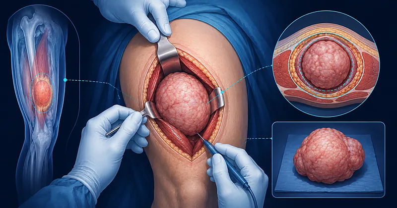
- 1Refer any deep soft tissue mass larger than 5 cm, or any growing superficial mass, to a sarcoma referral centre before biopsy → Pre-Op Workup
- 2Use image-guided core needle biopsy positioned along the planned resection axis — never an exploratory excisional biopsy → Biopsy Planning
- 3Aim for an R0 resection with a microscopically negative margin — a preserved anatomic barrier counts as a margin even when the distance is millimetric → Surgical Margins
- 4Excise the biopsy tract, surgical scar, and any drain site en bloc with the specimen → Surgical Margins
- 5Discuss preoperative radiotherapy for high-grade extremity tumours larger than 5 cm — lower long-term fibrosis at the cost of higher acute wound complications → Sequencing
- 6Consider neoadjuvant chemotherapy for high-risk localised disease — large, deep, high-grade tumours of chemosensitive histology → Sequencing
- 7Default to limb-sparing surgery whenever an R0 resection is achievable with acceptable function — amputation rates should fall below 5% in modern practice → Limb Salvage
- 8Plan reconstruction with plastic surgery before resection of any defect that may require flap coverage → Limb Salvage
- 9Submit oriented, inked specimens with the closest anticipated margin clearly identified for pathology → Surgical Margins
- 10Surveil with cross-sectional imaging of the primary site and CT chest every 3–6 months for the first 2–3 years — this is when most recurrences happen → Monitoring
Pre-Operative Workup and Biopsy Planning in Soft Tissue Sarcoma Surgery
The single most consequential decision in soft tissue sarcoma surgery is made at the moment of first consultation: who handles the biopsy. An unplanned excision performed at a non-specialist centre — the so-called “whoops procedure” — contaminates compartments, distorts anatomic landmarks, and frequently converts an otherwise limb-sparing case into a re-resection with worse oncologic and functional outcomes.
A complete pre-operative workup follows three sequential aims: anatomic delineation of the primary tumour, staging for metastatic disease, and tissue diagnosis with grading.
Refer any soft tissue mass that is deep to the fascia, larger than 5 cm, painful, growing, or recurrent to a sarcoma referral centre before any biopsy is taken.
Strong Rec High Evidence ESMO 2021 NCCN 2024Obtain MRI with contrast of the primary site as the initial imaging study for any extremity, trunk, or head-and-neck soft tissue mass — characterise size, depth, neurovascular involvement, and compartmental boundaries.
Strong Rec High Evidence NCCN 2024Use contrast-enhanced CT of the abdomen and pelvis as the first-line imaging study for any retroperitoneal or visceral soft tissue mass.
Strong Rec Moderate Evidence TARPSWG 2021Perform CT chest for staging in every histologically confirmed soft tissue sarcoma — the lung is by far the most common site of distant metastasis.
Strong Rec High Evidence NCCN 2024 ESMO 2021Perform image-guided core needle biopsy (minimum three cores, 14–16 gauge) as the standard approach to tissue diagnosis — sensitivity above 95% in experienced hands.
Strong Rec High Evidence ESMO 2021Place the biopsy tract along the line of the anticipated definitive incision so that the entire tract can be excised en bloc at surgery — coordinate with the operating surgeon before the biopsy if the radiologist is performing it.
Strong Rec Moderate Evidence NCCN 2024Do not use fine-needle aspiration as first-line for primary diagnosis — insufficient tissue for grading, subtyping, and molecular studies.
Against Moderate Evidence ESMO 2021Do not perform incisional or excisional biopsy as the initial tissue diagnosis in extremity sarcoma — reserve incisional biopsy for cases where core biopsy is non-diagnostic and only at the centre that will perform definitive surgery.
Against Moderate Evidence NCCN 2024Refer every histologically confirmed sarcoma to a dedicated multidisciplinary sarcoma board for review of imaging, pathology (including expert sarcoma pathology review), and treatment sequencing before surgery.
Strong Rec Moderate Evidence ESMO 2021Surgical Margins in Soft Tissue Sarcoma Surgery
Margin status is the single strongest modifiable predictor of local recurrence in soft tissue sarcoma surgery. The historical “wide local excision with 2 cm of normal tissue” rule oversimplifies a more nuanced reality: an intact anatomic barrier — periosteum, perineurium, major vessel adventitia, or fascia — functions as an oncologic margin even when the metric distance is small. Conversely, several centimetres of fat carry less oncologic value than a millimetre of preserved fascia.
Plan every soft tissue sarcoma surgery to achieve an R0 resection — microscopically negative margins in all directions.
Strong Rec High Evidence NCCN 2024 ESMO 2021Tailor margin width to anatomy and histology rather than applying a fixed numerical rule — 1–2 cm of soft tissue around the tumour where feasible, plus an intact fascial barrier wherever one is present.
Moderate Rec Moderate Evidence ESMO 2021Excise the biopsy tract, drain sites, prior scar tissue, and any seroma cavity en bloc with the specimen — treat any prior surgical contamination as part of the tumour.
Strong Rec Moderate Evidence NCCN 2024Do not violate the tumour pseudocapsule. Enucleation or “shelling out” results in microscopic residual disease and dramatically increases local recurrence.
Against High Evidence NCCN 2024Orient the specimen with sutures or anatomic landmarks and ink the closest anticipated margin before sending to pathology. Communicate directly with the sarcoma pathologist about anatomically tight areas.
Strong Rec Low Evidence NCCN 2024Document the closest anatomic margin in millimetres and the structure that constitutes that margin (fascia, periosteum, vessel adventitia) in the operative note — this directly informs radiation planning.
Strong Rec Low Evidence NCCN 2024Consider re-resection in patients with an R1 margin from prior unplanned excision — residual disease is found in roughly half of re-resection specimens and impacts both local control and survival.
Moderate Rec Moderate Evidence NCCN 2024Counsel patients with planned close margins near critical neurovascular structures that adjuvant radiotherapy will be required — preserve nerve, vessel, or bone rather than sacrifice function for an extra millimetre of fat.
Moderate Rec Moderate Evidence ESMO 2021Neoadjuvant Therapy Sequencing
For high-risk extremity sarcoma — large (>5 cm), deep, high-grade tumours — the question is rarely whether to add radiation or chemotherapy but when. Pre-operative radiotherapy delivers a smaller, more conformal field and lower late toxicity but is associated with substantially higher acute wound complications. Pre-operative chemotherapy can downsize borderline-resectable disease and treat occult micrometastases at the cost of delay and toxicity.
Offer preoperative radiotherapy (typically 50 Gy in 25 fractions) for high-grade, deep, extremity soft tissue sarcomas larger than 5 cm.
Strong Rec High Evidence O’Sullivan Trial 2002 NCCN 2024Counsel patients receiving preoperative radiotherapy to extremity sites about the higher risk of acute wound complications — roughly twice the rate seen with postoperative radiation, mainly affecting lower-extremity, posterior thigh tumours.
Strong Rec High Evidence O’Sullivan Trial 2002Schedule definitive surgery 4–6 weeks after completion of preoperative radiotherapy to allow acute inflammation to settle while staying within the window of maximum tumour response.
Moderate Rec Moderate Evidence ESMO 2021Consider neoadjuvant chemotherapy with an anthracycline-plus-ifosfamide regimen for patients with high-risk localised disease — large, deep, high-grade tumours of chemosensitive histology (synovial sarcoma, myxoid/round-cell liposarcoma, leiomyosarcoma, undifferentiated pleomorphic sarcoma, malignant peripheral nerve sheath tumour).
Moderate Rec Moderate Evidence ISG-STS 1001 ESMO 2021Reassess resectability with contrast-enhanced MRI of the primary site 4–6 weeks after the final cycle of neoadjuvant systemic therapy — size criteria alone underestimate response in some histologies.
Strong Rec Moderate Evidence NCCN 2024Consider isolated limb perfusion with melphalan and TNF-alpha for borderline-resectable extremity sarcoma where limb salvage would otherwise be impossible — only at experienced centres.
Conditional Rec Low Evidence ESMO 2021Add postoperative radiotherapy after R0 resection for any high-grade extremity tumour larger than 5 cm or any R1 resection — if preoperative radiation was not given.
Strong Rec High Evidence NCCN 2024 ESMO 2021Limb Salvage Decisions and Reconstruction
In modern soft tissue sarcoma surgery the default for extremity disease is limb-sparing resection, with amputation reserved for tumours that cannot be removed with adequate margins and acceptable function. The decision is not made by the surgeon alone — it is made by the multidisciplinary team, with the patient, and frequently with plastic surgery present in the operating room.
Default to limb-sparing surgery in extremity sarcoma whenever R0 margins can be achieved with an acceptable functional outcome — survival is equivalent to amputation when limb-sparing surgery is combined with radiation.
Strong Rec High Evidence NCCN 2024 ESMO 2021Refer every potential amputation candidate to multidisciplinary review before committing — consider whether neoadjuvant radiation, neoadjuvant chemotherapy, isolated limb perfusion, or planned reconstruction can convert the case to limb-sparing.
Strong Rec Moderate Evidence ESMO 2021Plan vascular reconstruction (interposition graft or bypass) at the same operation when major vessel resection is required — vessel involvement is no longer an automatic indication for amputation in experienced centres.
Moderate Rec Moderate Evidence NCCN 2024Involve plastic surgery preoperatively for any anticipated complex defect — pedicled or free flap, skin graft on irradiated tissue, or composite reconstruction. Mark flap territories before the oncologic resection begins.
Strong Rec Moderate Evidence NCCN 2024Consider intraoperative radiotherapy or brachytherapy in selected cases where the close margin abuts a critical structure that cannot be sacrificed — nerve, major vessel, or weight-bearing bone.
Conditional Rec Low Evidence NCCN 2024Reserve amputation for tumours where R0 limb-sparing resection is not achievable, where the residual limb would be non-functional, or where the patient prefers amputation after full counselling on alternatives.
Moderate Rec Moderate Evidence NCCN 2024Special Situations and Histology-Specific Considerations
Several anatomic locations and histologic subtypes demand approaches that diverge from the standard extremity algorithm.
For primary retroperitoneal sarcoma, perform a compartmental resection that includes adjacent organs at risk (kidney, colon, psoas, retroperitoneal fat) when needed to achieve R0 margins — piecemeal resection should be avoided.
Strong Rec Moderate Evidence TARPSWG 2021For desmoid-type fibromatosis, offer active surveillance as first-line for asymptomatic disease — spontaneous regression occurs in a significant minority and surgery carries a high recurrence rate.
Strong Rec Moderate Evidence ESMO 2021For myxoid liposarcoma, anticipate exceptional radiation sensitivity — consider lower-dose preoperative radiotherapy and smaller pre-op fields under multidisciplinary review.
Moderate Rec Moderate Evidence ESMO 2021Treat gastrointestinal stromal tumour (GIST) as a distinct entity — use neoadjuvant imatinib for marginally resectable disease and reserve compartmental dissection for adjacent organ involvement only.
Strong Rec High Evidence NCCN 2024For isolated locoregional recurrence after prior soft tissue sarcoma surgery, evaluate for re-resection — salvage surgery offers meaningful long-term survival in selected patients.
Moderate Rec Moderate Evidence NCCN 2024Consider metastasectomy for limited pulmonary metastatic disease in selected patients with prolonged disease-free interval and resectable lesions — long-term survival is achievable in a subset.
Conditional Rec Low Evidence ESMO 2021Clinical Decision Pathway
A practical, question-based approach to a newly identified soft tissue mass of concern. Work through the questions sequentially.
Practical Comparison Tables
Margin Terminology: What Each Designation Means in Practice
Different historical systems describe sarcoma margins. The table below maps each to the practical implication for the operating surgeon.
| Margin Designation | What It Means | Local Recurrence Implication | Next Step |
|---|---|---|---|
| R0 (clear) | No tumour at inked margin on microscopy | Lowest | Adjuvant RT if high-grade/>5 cm and no preop RT |
| R0 close | Clear but <1 mm or only fascial barrier | Slightly higher than R0 with wide tissue | Adjuvant RT routinely added |
| R1 | Microscopic tumour at the inked margin | Substantially elevated | Consider re-resection; if not feasible, adjuvant RT |
| R2 | Macroscopic residual disease | High | Re-resection essential; systemic disease reassessment |
| Enneking intralesional | Plane within the tumour itself | Very high — equivalent to R2 | Re-resection at sarcoma centre |
| Enneking marginal | Plane through the pseudocapsule | High — functionally equivalent to R1 | Re-resection or adjuvant RT |
Histology-Specific Considerations at a Glance
| Histology | Radiation Sensitivity | Chemotherapy Sensitivity | Surgical Pitfall |
|---|---|---|---|
| Undifferentiated pleomorphic sarcoma | Moderate | Moderate | Infiltrative borders — respect ≥1 cm margin where possible |
| Myxoid liposarcoma | High | Moderate | Round-cell component drives prognosis |
| Synovial sarcoma | Moderate | High | Often appears small radiographically; biopsy any indeterminate young-adult limb mass |
| Leiomyosarcoma | Low to moderate | Moderate | Vessel of origin involvement is common — plan for reconstruction |
| Well-differentiated liposarcoma | Low | Low | Margins are surgery-dependent; recurrence pattern is local |
| Malignant peripheral nerve sheath tumour | Low to moderate | Low to moderate | Plan proximal and distal nerve margins; counsel on functional deficit |
NCCN vs ESMO: Where the Two Frameworks Diverge
| Clinical Question | NCCN Approach | ESMO Approach | Where They Differ |
|---|---|---|---|
| Routine neoadjuvant chemotherapy | Reserved for high-risk localised disease in selected histologies | May be offered to high-risk extremity sarcoma after MDT review | ESMO slightly more permissive of histotype-tailored regimens |
| Adjuvant chemotherapy in extremity STS | Not routine; case-by-case | Consider in selected high-risk patients | Both frameworks emphasise individualised decisions |
| Retroperitoneal sarcoma radiotherapy | Preoperative RT may be considered case-by-case | Aligned with TARPSWG — selective use post-STRASS trial | STRASS trial reshaped both frameworks toward selective use |
| Surveillance frequency | Every 3–6 months for 2–3 years | Every 3–4 months for first 2–3 years | Largely aligned; differences are operational |
Monitoring and Surveillance
Most local recurrences and pulmonary metastases declare themselves within the first two to three years after definitive soft tissue sarcoma surgery. A structured surveillance plan increases the chance that recurrent disease is detected when it is still resectable.
| What to Check | Years 1–3 | Years 4–5 | Practical Tip |
|---|---|---|---|
| Clinical examination of primary site | Every 3–6 months | Every 6–12 months | Teach the patient to self-examine and report any new nodule |
| CT chest | Every 3–6 months | Every 6–12 months | Low-dose protocol where available to limit cumulative dose |
| MRI of primary site (extremity/trunk) | Every 6 months | Annually | Use same scanner protocol each time to allow direct comparison |
| CT abdomen/pelvis (retroperitoneal) | Every 3–6 months | Every 6–12 months | Pair with CT chest to capture both local and pulmonary recurrence |
| Functional assessment | At each visit | At each visit | Early physiotherapy improves long-term limb function after irradiated reconstruction |
Evidence in Context
What the major trials and frameworks actually show, and where ongoing controversy remains.
Pre- vs Post-Operative Radiation: The O’Sullivan Trial
The landmark Canadian randomised trial of preoperative versus postoperative external-beam radiation in extremity sarcoma showed similar local control between the two arms, with preoperative radiation associated with a roughly two-fold higher rate of acute wound complications — chiefly in lower extremity, posterior compartment tumours. Long-term follow-up favoured preoperative timing in terms of fibrosis, joint stiffness, and lymphoedema. The trial established preoperative radiation as a standard option, with the trade-off centred on acute wound morbidity versus late functional cost.
Neoadjuvant Chemotherapy: ISG-STS 1001
The Italian Sarcoma Group’s ISG-STS 1001 trial compared standard neoadjuvant epirubicin-plus-ifosfamide against histotype-tailored regimens in patients with high-risk localised extremity or trunk-wall soft tissue sarcoma. The standard arm performed at least as well as histotype-tailored therapy across the trial’s five subtypes, supporting anthracycline-plus-ifosfamide as a reasonable default neoadjuvant backbone for chemosensitive high-risk localised disease. The trial did not establish neoadjuvant chemotherapy as universally required — selection by histology, grade, size, and depth remains essential.
Retroperitoneal Sarcoma: STRASS and TARPSWG Consensus
The STRASS trial did not demonstrate an overall abdominal recurrence-free survival benefit for preoperative radiotherapy in primary retroperitoneal sarcoma at the trial-wide level, although subgroup signals for liposarcoma generated ongoing debate. Combined with the Trans-Atlantic RPS Working Group consensus, current practice emphasises high-quality compartmental surgery as the dominant determinant of local control, with radiation reserved for selected cases at experienced centres. The era of routine preoperative radiation for all retroperitoneal sarcoma has ended.
Where NCCN and ESMO Agree
Both frameworks converge on the centrality of multidisciplinary management, core needle biopsy with tract excision, R0 resection as the surgical goal, limb-sparing surgery with adjuvant or neoadjuvant radiation for high-risk extremity disease, and the surveillance schedule emphasising the first two to three postoperative years.
Ongoing Controversies
Several questions remain unresolved: the role of routine adjuvant chemotherapy in resected extremity sarcoma, the optimal margin width when no anatomic barrier is present, the place of immunotherapy in selected histologies, and how to predict which patients will benefit from histotype-tailored neoadjuvant regimens. Enrolment in clinical trials, where available, is encouraged for all high-risk and recurrent cases.
References
- 1.Gronchi A, Miah AB, Dei Tos AP, et al. Soft tissue and visceral sarcomas: ESMO-EURACAN-GENTURIS Clinical Practice Guidelines for diagnosis, treatment and follow-up. Ann Oncol. 2021;32(11):1348–1365. doi:10.1016/j.annonc.2021.07.006
- 2.O’Sullivan B, Davis AM, Turcotte R, et al. Preoperative versus postoperative radiotherapy in soft-tissue sarcoma of the limbs: a randomised trial. Lancet. 2002;359(9325):2235–2241. doi:10.1016/S0140-6736(02)09292-9
- 3.Gronchi A, Ferrari S, Quagliuolo V, et al. Histotype-tailored neoadjuvant chemotherapy versus standard chemotherapy in patients with high-risk soft-tissue sarcomas (ISG-STS 1001): an international, open-label, randomised, controlled, phase 3, multicentre trial. Lancet Oncol. 2017;18(6):812–822. doi:10.1016/S1470-2045(17)30334-0
- 4.Trans-Atlantic Retroperitoneal Sarcoma Working Group (TARPSWG). Management of primary retroperitoneal sarcoma (RPS) in the adult: an updated consensus approach from the Trans-Atlantic RPS Working Group. Ann Surg Oncol. 2021;28(12):7873–7888. doi:10.1245/s10434-021-09654-z
- 5.Bonvalot S, Gronchi A, Le PéchouxC, et al. Preoperative radiotherapy plus surgery versus surgery alone for patients with primary retroperitoneal sarcoma (EORTC-62092: STRASS): a multicentre, open-label, randomised, phase 3 trial. Lancet Oncol. 2020;21(10):1366–1377. doi:10.1016/S1470-2045(20)30446-0
- 6.von Mehren M, Kane JM, Agulnik M, et al. Soft Tissue Sarcoma, Version 2.2022, NCCN Clinical Practice Guidelines in Oncology. J Natl Compr Canc Netw. 2022;20(7):815–833. doi:10.6004/jnccn.2022.0035
How to Read the Evidence Tags
Every recommendation in this article carries simplified strength and evidence tags — Medaptly’s own interpretive shorthand to help the bedside clinician at a glance. For formal definitions, consult the original source documents listed in References.
Recommendation Strength
| Tag | What It Means |
|---|---|
| Strong Rec | High-quality evidence broadly supports this action. |
| Moderate Rec | The weight of evidence favours this action. |
| Conditional Rec | Benefit less certain — individualise. |
| Against | Evidence shows no benefit or potential harm. |
Evidence Quality
| Tag | What It Means |
|---|---|
| High Evidence | Multiple well-designed RCTs or high-quality meta-analyses. |
| Moderate Evidence | Single RCT or large observational studies. |
| Low Evidence | Expert consensus or small studies. |