
Ventral & Incisional Hernia Repair: A Mesh-and-Technique Guide
Clinical Practice Update — Mesh Selection, Plane Placement, and Repair Technique in Adults
This is an original clinical education article informed by current guidelines and evidence. See References below for source documents.
- Clinical Focus
- Mesh selection and repair technique for elective ventral hernia repair in adults
- Target Audience
- General and abdominal wall surgeons, surgical residents, surgical trainees
- Setting
- Elective operating theatre, ambulatory surgical centres, inpatient surgical units
- Source Evidence
- •European Hernia Society / Americas Hernia Society Guidelines on the Closure of Abdominal Wall Incisions and Ventral Hernia Management
- •Ventral Hernia Working Group (VHWG) Grading System for Surgical Site Occurrence Risk
- •STITCH Trial — Small Bites vs Large Bites for Midline Closure (Lancet, 2015)
- •Transversus Abdominis Release (TAR) Outcome Studies for Complex Reconstruction
Key Clinical Takeaways
Durable ventral hernia repair rests on three linked decisions: which mesh to implant, which anatomical plane to place it in, and how to close the fascia around it. Get any one wrong and recurrence climbs. The points below distil the evidence into rules you can apply when planning and executing an elective repair.
- 1Use mesh for almost every ventral hernia repair beyond a few millimetres — suture-only repair of an incisional hernia carries an unacceptable recurrence rate.
- 2Let defect width and contamination drive the plan more than any single classification label.
- 3Place mesh in the retromuscular (sublay) plane whenever the anatomy allows — it offers the best balance of durability and low complications.
- 4Choose lightweight-to-midweight permanent synthetic mesh for clean elective ventral hernia repair.
- 5Reserve intraperitoneal coated mesh for situations where no extraperitoneal plane is achievable.
- 6Aim for at least 5 cm of mesh overlap beyond the fascial edge in open retromuscular repair.
- 7Close the midline fascia over the mesh using a small-bites continuous technique with a slowly absorbable suture.
- 8Add a posterior component separation such as transversus abdominis release only when medial fascial advancement cannot otherwise be achieved.
- 9Optimise the patient before elective repair — smoking, glycaemic control, and obesity drive most preventable wound complications.
When Mesh Is Needed in Ventral Hernia Repair
The first decision in any ventral hernia repair is whether to reinforce the closure with mesh at all. For incisional hernias the answer is almost always yes: suture-only closure leaves recurrence rates several-fold higher than mesh reinforcement, and the gap widens with longer follow-up.
Use mesh reinforcement for essentially all incisional hernias and for primary ventral defects wider than roughly 1–2 cm. Suture-only repair may remain reasonable only for very small primary umbilical hernias in low-risk patients.
Strong Rec High Evidence EHS/AHS GuidelinesEvaluate for a planned, staged approach when the defect is very wide, loss of domain is present, or active contamination is expected. A definitive single-stage repair is not always the safest path.
Moderate Rec Moderate Evidence VHWGChoosing the Mesh Plane
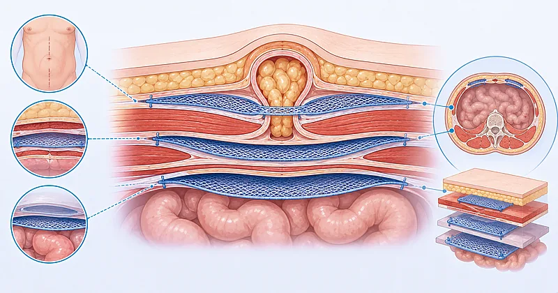
Where the mesh sits matters as much as what it is made of. The retromuscular plane, popularised by the Rives-Stoppa approach, places mesh behind the rectus muscle and in front of the posterior sheath, giving broad tissue ingrowth and keeping the prosthesis away from bowel.
Place mesh in the retromuscular (sublay) plane as the preferred position for most open ventral hernia repair. The Rives-Stoppa approach combines low recurrence with a low rate of mesh-related complications.
Strong Rec Moderate Evidence EHS/AHS GuidelinesAvoid routine use of the inlay (bridging) technique, where mesh spans the defect without fascial closure over it. Bridged repairs carry higher recurrence and bulging than reinforced closures.
Against Moderate Evidence EHS/AHS GuidelinesConsider the onlay plane (mesh above the anterior fascia) as a reasonable alternative when retromuscular dissection is not feasible, accepting a higher rate of seroma and wound issues from the wide subcutaneous dissection required.
Conditional Rec Low Evidence VHWGReserve the intraperitoneal onlay position for laparoscopic repair or when no extraperitoneal plane is achievable, and use only mesh with a visceral-side anti-adhesion barrier in that location.
Moderate Rec Moderate Evidence EHS/AHS GuidelinesMesh Planes at a Glance
| Plane (Surgeon’s Term) | Where Mesh Sits | Best Suited For | Watch Out For |
|---|---|---|---|
| Retromuscular (sublay) | Behind rectus, in front of posterior sheath | Most midline incisional hernias — preferred default | Longer dissection; respect the linea semilunaris |
| Onlay | On top of anterior fascia, under skin/fat | When sublay is not feasible; some open techniques | Seroma; wide flap raises wound-complication risk |
| Intraperitoneal (IPOM) | Inside peritoneal cavity against viscera | Laparoscopic repair; no extraperitoneal plane | Requires anti-adhesion barrier; adhesion risk |
| Inlay (bridging) | Sutured to defect edges, spanning the gap | Rarely advisable — last resort only | Highest recurrence and bulging; generally avoid |
Mesh Material Selection in Ventral Hernia Repair
Mesh material choice in ventral hernia repair turns on two questions: how clean is the field, and where will the mesh sit. In a clean elective case, a permanent synthetic mesh is the workhorse. Weight and pore size shape the long-term feel of the abdominal wall and the degree of foreign-body reaction.
Prescribe a lightweight or midweight large-pore permanent polypropylene mesh for clean (CDC class I) elective ventral hernia repair, matched to the wound classification of the case.
Strong Rec Moderate Evidence EHS/AHS GuidelinesDo not place uncoated polypropylene in direct contact with bowel. When mesh must sit intraperitoneally, select a product with a dedicated visceral-side anti-adhesion barrier.
Against Moderate Evidence EHS/AHS GuidelinesConsider biologic or biosynthetic mesh in clean-contaminated or contaminated fields only after weighing cost against an uncertain durability benefit; permanent synthetic mesh performs acceptably in many such cases.
Conditional Rec Low Evidence VHWGEnsure mesh overlap of at least 5 cm beyond the fascial defect edge in open retromuscular repair, and a wide circumferential overlap in laparoscopic repair, to distribute load and reduce edge recurrence.
Strong Rec Moderate Evidence EHS/AHS GuidelinesThink in three field categories rather than memorising product names. In a clean field, permanent synthetic is standard. In a clean-contaminated field, permanent synthetic remains reasonable with sound technique and antibiotics. In a frankly contaminated or dirty field, the decision becomes individualised — some surgeons stage the repair, some use biosynthetic absorbable scaffolds, and the evidence does not crown a single winner.
Fascial Closure and Component Separation
Restoring the midline so the fascia meets over the mesh is the goal of modern repair. How that closure is sewn has its own evidence base: the STITCH trial showed that a small-bites technique for elective midline laparotomy closure reduces incisional hernia formation compared with traditional large bites.
Close the midline fascia over the mesh whenever achievable, converting a bridged repair into a reinforced one. Fascial reapproximation restores the linea alba and lowers recurrence.
Strong Rec Moderate Evidence EHS/AHS GuidelinesPerform fascial closure with a continuous, slowly absorbable suture using small bites (roughly 5 mm of fascia, 5 mm apart) and a suture-length-to-wound-length ratio of at least 4 to 1.
Strong Rec High Evidence STITCH 2015Consider transversus abdominis release to gain medial fascial advancement in wide defects where the posterior sheath cannot otherwise be closed, after less extensive options have been exhausted.
Moderate Rec Moderate Evidence TAR StudiesAvoid routine use of anterior component separation with wide lipocutaneous flaps when a posterior release or sublay repair would achieve closure, given the higher wound-complication burden of large skin flaps.
Conditional Rec Low Evidence VHWGClinical Decision Pathway
A practical, question-based route through mesh and technique decisions for an elective ventral hernia repair. Work through the questions in order.
Matching Mesh and Technique to the Clinical Scenario
This scenario-led table pairs common presentations with a sensible default plane, material, and closure approach. Treat it as a starting point to be tailored to the individual patient and your own operative judgement.
| Clinical Scenario | Default Plane | Mesh Material | Closure Approach | Practical Tip |
|---|---|---|---|---|
| Small primary umbilical hernia, clean | Pre-peritoneal / sublay | Small flat synthetic mesh | Primary closure over mesh | Even small defects do better with mesh than suture alone |
| Midline incisional hernia, moderate width, clean | Retromuscular (Rives-Stoppa) | Lightweight large-pore polypropylene | Small-bites continuous | Aim for ≥5 cm overlap on all sides |
| Wide defect, fascia will not meet | Retromuscular + posterior release | Midweight synthetic | TAR then small-bites closure | Release only as far as needed for tension-free midline |
| Laparoscopic repair, no extraperitoneal plane | Intraperitoneal (IPOM) | Barrier-coated mesh | Defect closure plus wide overlap | Orient the barrier toward the viscera |
| Contaminated field | Sublay where feasible | Individualised — synthetic vs biosynthetic | Tension-free closure, consider staging | Source control and antibiotics come first |
Monitoring and Follow-Up
Postoperative surveillance focuses on early wound problems and on the longer arc of recurrence. The table below frames what to watch, when, and the mistakes that catch people out.
| What to Watch | When | What to Look For | Common Pitfall |
|---|---|---|---|
| Wound and surgical site occurrence | First 30 days | Erythema, drainage, seroma, surgical site occurrence requiring intervention | Aspirating a stable seroma unnecessarily and seeding infection |
| Seroma | Weeks 1–6 | Most resolve spontaneously; persistent or symptomatic collections need review | Mistaking an expected post-onlay seroma for recurrence |
| Recurrence | 6 months and beyond | New bulge, palpable fascial edge, symptoms on straining | Stopping follow-up too early — recurrence can be late |
| Chronic pain or mesh sensation | Ongoing | Persistent focal pain, restriction, or discomfort over the mesh | Attributing all pain to the mesh without excluding other causes |
Monitor the wound through the first month for early surgical site occurrence, and counsel patients that a soft fluctuant swelling in the first weeks is often a self-limiting seroma rather than recurrence.
Moderate Rec Low Evidence VHWGReassess at intervals beyond six months for late recurrence, since recurrences after ventral hernia repair can appear years after an apparently successful operation.
Moderate Rec Low Evidence EHS/AHS GuidelinesEvidence in Context
What the trials and guidelines show, where they align, and where genuine uncertainty remains.
Why Mesh Beats Suture Repair
Randomised and registry data consistently show that mesh reinforcement substantially lowers recurrence after incisional hernia repair compared with suture-only closure, and the advantage persists or grows over long follow-up. This is the most settled question in the field.
Small Bites and the STITCH Trial
The STITCH trial randomised elective midline laparotomy closures to small versus large bites and found fewer incisional hernias at one year in the small-bites group. The technique has since been widely adopted for both primary closure and closure over mesh in ventral hernia repair.
Mesh in Contaminated Fields: Unsettled Ground
The choice between permanent synthetic, biosynthetic absorbable, and biologic mesh in contaminated fields remains debated. Synthetic mesh performs better in such fields than older teaching suggested, while biologic mesh has not delivered the durability once hoped, and head-to-head evidence is limited.
Posterior vs Anterior Component Separation
Posterior approaches such as transversus abdominis release avoid the large lipocutaneous flaps of classic anterior separation, and observational series report lower wound-complication rates. Comparative trial evidence is still maturing, so technique choice often reflects defect anatomy and surgeon experience.
References
- 1.Muysoms FE, Antoniou SA, Bury K, et al. European Hernia Society guidelines on the closure of abdominal wall incisions. Hernia. 2015;19(1):1–24. doi:10.1007/s10029-014-1342-5
- 2.Deerenberg EB, Harlaar JJ, Steyerberg EW, et al. Small bites versus large bites for closure of abdominal midline incisions (STITCH): a randomised controlled trial. Lancet. 2015;386(10000):1254–1260. doi:10.1016/S0140-6736(15)60459-7
- 3.Novitsky YW, Elliott HL, Orenstein SB, Rosen MJ. Transversus abdominis muscle release: a novel approach to posterior component separation during complex abdominal wall reconstruction. Am J Surg. 2012;204(5):709–716. doi:10.1016/j.amjsurg.2012.02.008
- 4.Breuing K, Butler CE, Ferzoco S, et al. Incisional ventral hernias: review of the literature and recommendations regarding the grading and technique of repair. Surgery. 2010;148(3):544–558. doi:10.1016/j.surg.2010.01.008
- 5.Liang MK, Holihan JL, Itani K, et al. Ventral Hernia Management: Expert Consensus Guided by Systematic Review. Ann Surg. 2017;265(1):80–89. doi:10.1097/SLA.0000000000001701
How to Read the Evidence Tags
Each recommendation carries a strength tag and an evidence-quality tag — Medaptly’s own simplified interpretations, not any guideline body’s classification system. For full grading definitions, consult the original source documents in the References.
Recommendation Strength
| Tag | What It Means |
|---|---|
| Strong Rec | High-quality evidence broadly supports this action. |
| Moderate Rec | The weight of evidence favours this action. |
| Conditional Rec | The benefit is less certain — individualise. |
| Against | Evidence shows no benefit or potential harm. |
Evidence Quality
| Tag | What It Means |
|---|---|
| High Evidence | Multiple well-designed RCTs or high-quality meta-analyses. |
| Moderate Evidence | Single RCT or large observational studies. |
| Low Evidence | Expert consensus or small studies. |