
Pediatric UTI Imaging: When Ultrasound, VCUG, and DMSA Change Management
Renal ultrasound, VCUG, and DMSA each answer a different question after a first febrile urinary tract infection — and only some of those answers change what you do next.
One-Minute Takeaway
- Pediatric UTI imaging draws on strong evidence — randomized trials and a Cochrane diagnostic meta-analysis — yet after a first febrile UTI, most imaging does not change management.
- Renal and bladder ultrasound is normal in roughly 75% to 88% of children, and its abnormalities rarely alter treatment; its real value lies in detecting obstruction, not reflux.
- Ultrasound is an insensitive screen for reflux (pooled sensitivity about 44% for any VUR, 59% for high-grade), so a normal scan cannot exclude it.
- A negative DMSA scan makes high-grade reflux very unlikely (under 1% probability), but its low specificity refers many children to avoidable VCUG.
- Prophylaxis for reflux reduces recurrences by roughly half (relative risk 0.55) but does not reduce scarring — limiting the payoff of finding low-grade reflux.
Why Pediatric UTI Imaging Matters
Urinary tract infection is among the most common serious bacterial infections in young children. Roughly one-third of children presenting with a febrile UTI have vesicoureteral reflux (VUR), which has long been linked to recurrent infection and renal scarring.
For decades, this association drove an aggressive reflex: image every child, find the reflux, and treat it. Renal-bladder ultrasound (RBUS) and voiding cystourethrography (VCUG) were performed routinely, and reflux was managed with prolonged antimicrobial prophylaxis or surgery.
That model has been steadily challenged. The core question for pediatric UTI imaging is no longer “can we find an abnormality” but “does finding it change what we do.” Each available test detects something different, at a different cost in radiation, invasiveness, and money.
When high-grade reflux or an obstructive anomaly is present, imaging genuinely shapes care. The difficulty is that these findings are uncommon after a first infection, while the tests used to hunt for them carry real burdens for every child imaged.
The stakes are not trivial. Reflux nephropathy — scarring attributed to reflux and recurrent infection — has historically been linked to hypertension and, in a minority of children, chronic kidney disease. Those long-term risks are precisely why pediatric UTI imaging was once pursued so aggressively, and why any shift toward less testing must be justified by outcomes rather than convenience.
The Clinical Problem in Pediatric UTI Imaging
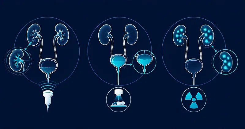
Three tools dominate the workup, and each targets a different structure. Ultrasound assesses anatomy. VCUG demonstrates reflux and grades its severity. The DMSA scan shows renal parenchymal involvement and later scarring.
Two philosophies organize their use. The “bottom-up” approach starts from the bladder with VCUG to find reflux. The “top-down” approach starts from the kidney with an acute DMSA scan to find parenchymal inflammation, then applies VCUG selectively to children with abnormal scans.
The pivotal shift came when guidance stopped recommending routine VCUG after a first febrile UTI, retaining ultrasound as the first-line study. The rationale rested on three observations that reframed the whole question.
- High-grade reflux is uncommon after a first febrile UTI, so most VCUGs return low-grade or negative results.
- Antimicrobial prophylaxis, the main intervention triggered by finding reflux, has a modest effect on recurrence and none on scarring.
- Reliable urinalysis and culture at future febrile illnesses can catch recurrences without pre-emptive invasive imaging.
Against that backdrop, “changing management” becomes a high bar. A test earns its place only when its result plausibly redirects treatment toward something that improves outcomes — not merely when it labels a child as having reflux.
Diagnostic thresholds shape the entire pathway. A UTI is defined by pyuria together with a meaningful single-organism culture, and only a confirmed infection should trigger pediatric UTI imaging at all. Over-diagnosis at the front door inflates the number of children sent for tests whose results were never likely to change their care.
Available Approaches
The table below summarizes the verified evidence comparing the imaging modalities and the interventions they trigger. Every figure was checked against a primary source, and each reference resolves to the cited paper.
| Study / Source | Design | N | Focus | Key result | Conf. |
|---|---|---|---|---|---|
| Shaikh 2016 (Cochrane) | Diagnostic systematic review & meta-analysis | 19–20 studies | RBUS vs DMSA to screen for VUR | RBUS sensitivity 0.44 (95% CI 0.34–0.54) for any VUR; DMSA sensitivity 0.93 (95% CI 0.77–0.98) for high-grade VUR; negative DMSA → <1% probability of high-grade VUR | V |
| Nelson 2014 (Pediatrics) | Retrospective diagnostic cohort | 1,203 | RBUS test characteristics for VCUG findings | RBUS sensitivity 5–28% for any abnormal VCUG finding; PPV 13–24% and NPV 95–96% for VUR grade >III; RBUS “poor screening test” | V |
| Hoberman 2003 (NEJM) | Prospective trial | 309 | Whether imaging alters management | Ultrasound normal in 88%; abnormalities did not modify management; VUR in 39%, of which 96% was grade I–III; abnormal (acute) DMSA at presentation in 9.5% | V |
| Fouzas 2010 (Pediatrics) | Prospective diagnostic cohort | 296 | Acute DMSA to reveal VUR | Acute DMSA for dilating VUR: AUC 0.68, negative likelihood ratio 0.46; concluded DMSA should not replace VCUG | V |
| RIVUR 2014 (NEJM) | Randomized controlled trial | 607 | Prophylaxis vs placebo in children with VUR | Recurrence relative risk 0.55 (95% CI 0.38–0.78); no significant reduction in renal scarring (11.9% vs 10.2%) | V |
Renal and Bladder Ultrasound (RBUS)
Ultrasound is noninvasive, radiation-free, and widely available, which makes it the natural first study. It reliably identifies gross structural problems: hydronephrosis, duplex systems, ureteroceles, and other congenital anomalies of the kidney and urinary tract.
Its weakness is reflux. In the Cochrane meta-analysis, pooled sensitivity was only 0.44 (95% CI 0.34–0.54) for any VUR and 0.59 (95% CI 0.45–0.72) for high-grade VUR. A large diagnostic cohort found sensitivity as low as 5% to 28% for detecting any abnormality on cystography.
Strong evidence supports a narrow reading: a normal ultrasound cannot exclude reflux, but it can reassure that a treatable obstructive lesion is unlikely. Ultrasound changes management chiefly when it uncovers anatomy that itself warrants intervention or further targeted imaging.
Two practical caveats matter. A reassuring antenatal ultrasound lowers the pre-test probability of a major structural anomaly, and the prevalence of dilated uropathy detected after infection has fallen over time. Timing also matters: a scan performed during acute illness can show pelvic dilation from inflammation that mimics true hydronephrosis, so early findings are sometimes read cautiously.
Voiding Cystourethrography (VCUG) — the bottom-up target
VCUG is the reference standard for diagnosing reflux and assigning its grade. That precision comes at a price: bladder catheterization, ionizing radiation, and considerable distress for child and family.
The problem is what it finds. In the landmark prospective series, reflux was present in 39% of children, but 96% of it was low-grade (grades I–III). Dilating high-grade reflux — the kind that plausibly alters care — is uncommon after a first infection.
VCUG changes management mainly when it reveals high-grade (IV–V) reflux, which is a candidate for closer surveillance, prophylaxis, or surgical referral. For the far larger group with low-grade reflux, the result rarely redirects treatment. Current practice reserves VCUG for an abnormal ultrasound, atypical features, or a second febrile UTI.
The natural history reinforces this restraint. Low-grade reflux resolves spontaneously in many children as they grow, so a diagnosis often carries no durable consequence. Weighed against catheterization, ionizing radiation to the pelvis, and the distress the study causes, routine cystography after a first infection asks a great deal for information that seldom alters the plan.
DMSA Scintigraphy — the top-down target
DMSA is the most sensitive study for acute pyelonephritis and for established renal scarring. In the top-down strategy, an acute DMSA identifies parenchymal involvement, and only children with abnormal scans proceed to VCUG.
The screening logic is genuinely attractive on one axis. A negative DMSA scan makes high-grade reflux very unlikely, with a reported probability under 1%. That negative predictive value could, in principle, spare many children a catheter-based study.
The offsetting problems are radiation, cost, and low specificity. A prospective cohort found acute DMSA only modestly discriminating for dilating reflux (AUC 0.68), and its authors concluded the scan should not replace VCUG. Positive scans are common, so many children still face downstream cystography.
Timing separates two distinct uses. An acute DMSA maps active pyelonephritis, while a scan repeated months later distinguishes transient inflammation from permanent scarring. Late scarring carries prognostic weight, but detecting it does not by itself dictate an intervention — making its role in pediatric UTI imaging more about risk stratification than about an immediate change in management.
Comparative Analysis
The three modalities are not interchangeable; they answer different clinical questions. Read together, the evidence points to when each earns its place after a first febrile UTI.
Ultrasound changes management when it finds obstruction or a major structural anomaly — uncommon, but consequential when present. It is a poor detector of reflux and should not be relied on to exclude it.
VCUG changes management when it demonstrates high-grade reflux. Because such reflux is infrequent after a first infection and most detected reflux is low-grade, routine use exposes many children to an invasive test for little gain.
DMSA changes management when parenchymal involvement or scarring alters prognosis, or when a normal scan is used to withhold VCUG. A retrospective comparison of five imaging strategies found the top-down approach the most sensitive for detecting reflux (76%) and scarring (100%), but the least specific of the strategies compared and among the most costly and radiation-heavy.
The unifying reason the gains stay small is downstream. Because prophylaxis reduces recurrence but not scarring, and because high-grade reflux is uncommon, detecting reflux seldom leads to an intervention that changes long-term outcomes for the average child.
Prophylaxis also carries a cost that weighs against reflexive VCUG. In the randomized trial, among children whose first recurrence was caused by E. coli, resistance to trimethoprim-sulfamethoxazole reached 63% in the prophylaxis group versus 19% with placebo. Selecting more children for prophylaxis by finding more reflux therefore trades a modest recurrence benefit for more resistant organisms.
A useful frame is to ask what each positive result would trigger. If the answer is “an intervention that improves outcomes” — relieving obstruction, or surveilling high-grade reflux — the test is worth ordering. If the answer is “a label without a change in plan,” the test mainly adds cost, radiation, or discomfort.
Quality and Consistency of the Evidence
Evidence strength varies sharply by question. The diagnostic-accuracy data are robust and consistent; the outcome data behind management decisions are thinner.
- Strong — Ultrasound is insensitive for reflux. A large Cochrane meta-analysis and a multi-thousand-encounter cohort agree that a normal ultrasound cannot exclude VUR.
- Strong — Prophylaxis reduces recurrence but not scarring. A randomized placebo-controlled trial and supporting trials show a modest recurrence benefit with no scarring benefit.
- Moderate — Imaging rarely changes acute management. A prospective series showed ultrasound abnormalities did not alter treatment, and guideline bodies have reaffirmed selective imaging.
- Moderate — DMSA and the top-down approach have real but heterogeneous accuracy for significant reflux, with wide confidence intervals across studies of differing design.
- Limited — Whether any single imaging strategy improves long-term renal outcomes is untested by randomized trials with hard endpoints such as chronic kidney disease.
What the Evidence Does Not Show
Several claims often attached to post-UTI imaging are not supported by the current evidence and should be stated plainly.
The literature does not show that detecting and treating low-grade reflux improves long-term renal function. It does not show that any imaging strategy reduces chronic kidney disease or mortality, because no strategy has been tested against those endpoints in a randomized trial.
The evidence also does not establish that the top-down approach improves patient outcomes relative to selective imaging; the comparisons available address test characteristics, cost, and radiation, not downstream health.
Two practical questions remain open. The optimal approach for infants under two months is not settled, since much of the guideline evidence was generated in older infants. And whether the timing of ultrasound, acute versus delayed, meaningfully changes its yield is not firmly resolved.
Cost-effectiveness is likewise unsettled. Analyses comparing strategies can weigh dollars, radiation, and detection rates, but they cannot capture a health benefit the outcome trials have not demonstrated. Until a strategy is shown to move hard renal endpoints, the choice between approaches rests on how a family and clinician weigh a small, uncertain diagnostic yield against concrete procedural burdens.
Practical Implications
The following reflects what the evidence supports, not a directive for any individual child; clinical judgment and local guidance govern care.
- Ultrasound after a first febrile UTI is reasonable and low-burden. Its principal value is identifying obstruction or major structural anomalies, not screening for reflux.
- VCUG is not a routine first study. The evidence supports reserving it for an abnormal ultrasound, atypical or complex presentations, or a recurrent febrile UTI.
- A negative DMSA can lower suspicion for high-grade reflux, but routine DMSA or a universal top-down protocol adds radiation and cost for most children.
- Because prophylaxis reduces recurrence but not scarring, finding low-grade reflux seldom warrants a change in management.
- Reliable urinalysis and culture at future febrile illnesses can substitute for pre-emptive invasive imaging in many children.
- Individual risk — recurrent infection, bladder and bowel dysfunction, or a strong family history — should weigh toward more thorough evaluation.
- When imaging is deferred, a clear plan for prompt evaluation of the next febrile illness is the safeguard that makes watchful waiting reasonable.
Evidence Grade and Bottom Line
Bottom line
The core strategy — ultrasound first, VCUG selectively — rests on moderate-to-high-quality evidence. The case for the top-down DMSA approach is moderate and turns on local priorities around radiation and cost.
Imaging changes management for a minority of children after a first febrile UTI: ultrasound when it reveals obstruction, VCUG when it reveals high-grade reflux, and DMSA when parenchymal involvement alters prognosis or gates cystography. For most children, extensive imaging does not change what you do — and that, not the presence of reflux, is the finding that should guide the workup.
Article Information and References
- Shaikh N, Spingarn RB, Hum SW. Dimercaptosuccinic acid scan or ultrasound in screening for vesicoureteral reflux among children with urinary tract infections. Cochrane Database Syst Rev. 2016;7(7):CD010657. DOI: https://doi.org/10.1002/14651858.CD010657.pub2
- Mantadakis E, Vouloumanou EK, Georgantzi GG, Tsalkidis A, Chatzimichael A, Falagas ME. Acute Tc-99m DMSA scan for identifying dilating vesicoureteral reflux in children: a meta-analysis. Pediatrics. 2011;128(1):e169–e179. DOI: https://doi.org/10.1542/peds.2010-3460
- La Scola C, De Mutiis C, Hewitt IK, et al. Different guidelines for imaging after first UTI in febrile infants: yield, cost, and radiation. Pediatrics. 2013;131(3):e665–e671. DOI: https://doi.org/10.1542/peds.2012-0164
- Nelson CP, Johnson EK, Logvinenko T, Chow JS. Ultrasound as a screening test for genitourinary anomalies in children with UTI. Pediatrics. 2014;133(3):e394–e403. DOI: https://doi.org/10.1542/peds.2013-2109
- Fouzas S, Krikelli E, Vassilakos P, Gkentzi D, Papanastasiou DA, Salakos C. DMSA scan for revealing vesicoureteral reflux in young children with urinary tract infection. Pediatrics. 2010;126(3):e513–e519. DOI: https://doi.org/10.1542/peds.2009-3635
- Hoberman A, Charron M, Hickey RW, Baskin M, Kearney DH, Wald ER. Imaging studies after a first febrile urinary tract infection in young children. N Engl J Med. 2003;348(3):195–202. DOI: https://doi.org/10.1056/NEJMoa021698
- RIVUR Trial Investigators; Hoberman A, Greenfield SP, Mattoo TK, et al. Antimicrobial prophylaxis for children with vesicoureteral reflux. N Engl J Med. 2014;370(25):2367–2376. DOI: https://doi.org/10.1056/NEJMoa1401811
- Roberts KB; Subcommittee on Urinary Tract Infection, Steering Committee on Quality Improvement and Management. Urinary tract infection: clinical practice guideline for the diagnosis and management of the initial UTI in febrile infants and children 2 to 24 months. Pediatrics. 2011;128(3):595–610. DOI: https://doi.org/10.1542/peds.2011-1330
- Subcommittee on Urinary Tract Infection. Reaffirmation of AAP Clinical Practice Guideline: The Diagnosis and Management of the Initial Urinary Tract Infection in Febrile Infants and Young Children 2–24 Months of Age. Pediatrics. 2016;138(6):e20163026. DOI: https://doi.org/10.1542/peds.2016-3026
- Mattoo TK, Shaikh N, Nelson CP. Contemporary Management of Urinary Tract Infection in Children. Pediatrics. 2021;147(2):e2020012138. DOI: https://doi.org/10.1542/peds.2020-012138